. 2004 Sep;186(18):6003-14.
doi: 10.1128/JB.186.18.6003-6014.2004.

DegU-P represses expression of the motility fla-che operon in Bacillus subtilis

Affiliations

DegU-P represses expression of the motility fla-che operon in Bacillus subtilis

Giuseppe Amati et al. J Bacteriol. 2004 Sep.

Abstract

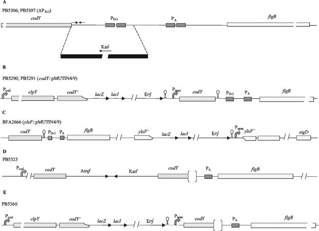
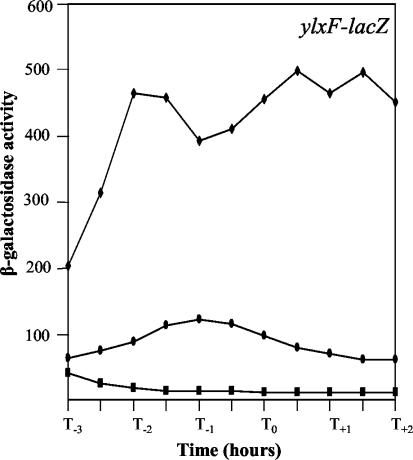
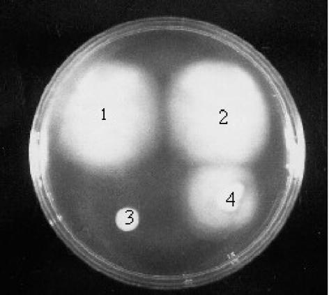
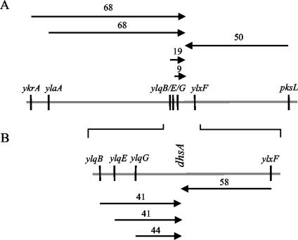
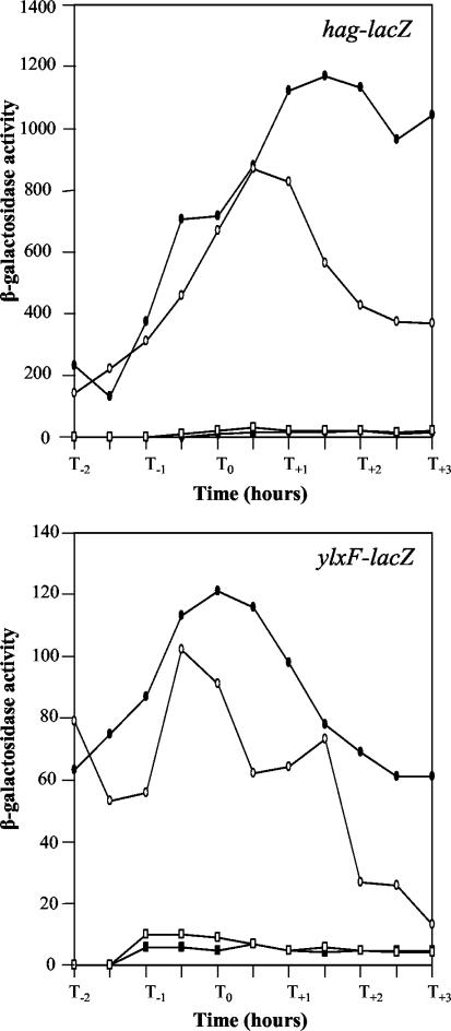
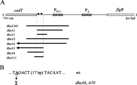
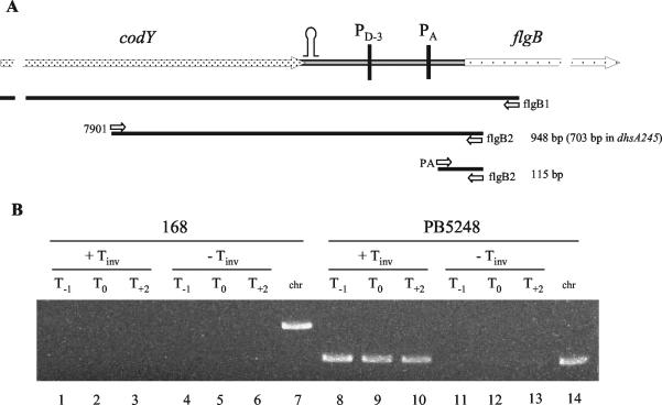
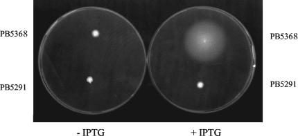
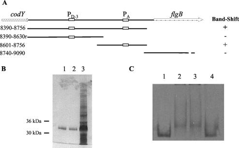
Bacillus subtilis implements several adaptive strategies to cope with nutrient limitation experienced at the end of exponential growth. The DegS-DegU two-component system is part of the network involved in the regulation of postexponential responses, such as competence development, the production of exoenzymes, and motility. The degU32(Hy) mutation extends the half-life of the phosphorylated form of DegU (DegU-P); this in turn increases the production of alkaline protease, levan-sucrase, and other exoenzymes and inhibits motility and the production of flagella. The expression of the flagellum-specific sigma factor SigD, of the flagellin gene hag, and of the fla-che operon is strongly reduced in a degU32(Hy) genetic background. To investigate the mechanism of action of DegU-P on motility, we isolated mutants of degU32(Hy) that completely suppressed the motility deficiency. The mutations were genetically mapped and characterized by PCR and sequencing. Most of the mutations were found to delete a transcriptional termination signal upstream of the main flagellar operon, fla-che, thus allowing transcriptional readthrough from the cod operon. Two additional mutations improved the sigmaA-dependent promoter sequence of the fla-che operon. Using an electrophoretic mobility shift assay, we have demonstrated that purified DegU binds specifically to the PA promoter region of the fla-che operon. The data suggest that DegU represses transcription of the fla-che operon, and they indicate a central role of the operon in regulating the synthesis and assembly of flagella.


Figures

FIG. 1.
FIG. 1.
Schematic presentation of construction of B. subtilis mutant strains. (A) Deletion of PD-3 promoter sequence upstream of fla-che operon. flgB is the first gene of the operon. The region between the end of the codY gene and the PA promoter was replaced with the kanamycin resistance determinant of plasmid pJM114 by a double-crossover event. (B) Placement of codY gene under control of the IPTG-inducible Pspac promoter. A chromosomal fragment corresponding to the end of the clpY gene and to one-third of the codY gene was amplified by PCR and cloned into pMutin4. The pMutin4 derivative was inserted into the chromosome by a single crossover event (Campbell-type integration), thus placing the codY gene under the control of Pspac. (C) Construction of ylxF-lacZ transcriptional fusion. The ylxF gene of the fla-che operon was disrupted with pMutin4 by a Campbell-type integration, placing the lacZ reporter gene under the control of the Pspac promoter. (D) Insertion of the pJM114 plasmid sequence upstream of the dhsA4 deletion mutation. The codY coding sequence was amplified and cloned into plasmid pJM114. The plasmid derivative was inserted into the chromosome of strain PB5317 (dhsA4) by a Campbell-type event. (E) Placement of fla-che operon gene under control of Pspac promoter. The codY gene under the control of Pspac was combined with a deletion of the transcription terminator dhsA245. The deletion is represented by an empty space between the two brackets. The densely dotted boxes refer to the clpY and codY genes, which are part of the cod operon. The fla-che operon genes (flgB, ylxF, and sigD) are shown as sparsely dotted boxes. The stem-loops indicate transcription termination signals. lacI, E. coli lacI gene; Ampr, ampicillin resistance marker; Kanr, kanamycin resistance marker; Eryr, erythromycin resistance marker.
FIG. 2.
FIG. 2.
Analysis of fla-che operon expression in different genetic backgrounds. The ylxF-lacZ fusion schematically shown in Fig. 1C was used to monitor the expression of the fla-che operon in different B. subtilis strains. ylxF is the ninth open reading frame of the operon. The strains were BFA 2666 (wild type; solid circles) PB5279 [dehU32(Hy); solid squares], and PB5284 [degU32(Hy) dhsA245; solid diamonds]. The β-galactosidase specific activity is expressed in Miller units per milligram of protein. Zero time (T0) indicates the transition point between the exponential and stationary phases of growth.
FIG. 3.
FIG. 3.
Motility of B. subtilis strains. Overnight colonies were transferred to a motility plate and incubated at 37°C overnight. 1, strain PB5248 [degU32(Hy) ΔdhsA245]; 2, strain PB5327 (ΔdhsA245 ΔdegSU); 3, strain PB5213 [degU32(Hy)]; 4, strain PB168 (wild type).
FIG. 4.
FIG. 4.
Genetic map of dhsA245 mutation. (A) Transduction crosses; (B) transformation crosses. The arrows are based on the selected marker and point to the nonselected marker. Donor strains had an erythromycin resistance determinant present in the indicated gene that was used as a selective marker. After selection, the transductants (transformants) were screened for motility. Distances are expressed as percentages of cotransduction (cotransformation). The gray line in panel A represents the region between 130° and 155° on the genetic map.
FIG. 5.
FIG. 5.
Deletion of the PD-3 promoter does not affect hag gene (A) and fla-che operon (B) expression. hag-lacZ (9) and ylxF-lacZ (Fig. 1C) transcriptional fusions were used to monitor the expression of the flagellin hag gene and the fla-che operon, respectively. The strains were PB5128 and BFA2666 (wild type; filled circles), PB5324 and PB5328 (ΔPD-3; open circles), PB5267 and PB5279 [degU32(Hy); open squares], and PB5325 and PB5329 [degU32(Hy) ΔPD-3; filled squares].
FIG. 6.
FIG. 6.
Molecular analysis of codY-flgB chromosomal region. Chromosomal DNAs from B. subtilis strains were PCR amplified with primers 7541 and flgB (Table 2 and Fig. 7) and were analyzed by agarose gel (0.6%) electrophoresis. Lanes 3 and 13, molecular weight standard (SPP1 phage DNA digested with EcoRI). The DNA samples were as follows: lane 1, PB5213 (wild type); lane 2, PB5248 (Δdhs245); lane 4, PB5314 (ΔdhsA1); lane 5, PB5315 (ΔdhsA2); lane 6, PB5316 (ΔdhsA3); lane 7, PB5317 (ΔdhsA4); lane 8, PB5318 (ΔdhsA5); lane 9, PB5319 (dhsA6); lane 10, PB5320 (ΔdhsA8); lane 11, PB5321 (dhsA10); lane 12, PB5322 (ΔdhsA11).
FIG. 7.
FIG. 7.
Mutations of dhs locus. (A) Schematic physical map of codY-flgB region of B. subtilis chromosome. The open arrows labeled 7541 and flgB indicate the positions of the oligonucleotide primers used to amplify the codY-flgB region (Fig. 6). The positions and extension of eight dhs deletion mutations are indicated by the solid bars below the physical map. Deletions Δdhs4 and Δdhs5 extend further into the cod operon and are represented by arrows pointing to the left. (B) Sequence of the σA-dependent promoter PA and of the dhsA6 and dhsA10 point mutations.
FIG. 8.
FIG. 8.
RT-PCR analysis of transcriptional readthrough. (A) Diagram of codY-flgB region. PD-3 and PA indicate the σD-dependent and σA-dependent promoters, respectively. The stem-loop indicates the transcription terminator at the end of the cod operon. The open arrows indicate the oligonucleotides used to prime RT (flgB1) and to amplify the cDNA products (7901 and flgB2; PA and flgB2). The calculated sizes of the PCR products are reported to the right. (B) Ethidium bromide-stained 1% agarose gel of RT-PCR and PCR products. RT reactions with the RNAs obtained from strains PB168 and PB5248 (as indicated) were performed with the flgB1 primer. The cDNAs were amplified with primers 7901 and flgB1. T0 refers to the time of transition between the exponential growth and stationary phases; T−1 and T2 refer to 1 h earlier and 2 h later, respectively. Lanes 1 to 6, the template for RT-PCRs was RNA from PB168 (wild type); lanes 1 to 3, RT-PCRs with reverse transcriptase; lanes 4 to 6, reactions in the absence of reverse transcriptase. Lane 7, PCR with chromosomal DNA from strain PB168 as a template and with primers 7901 and flgB2. Lanes 8 to 13, RT-PCRs with RNA from strain PB5248 (ΔdhsA245) as a template; lanes 8 to 10, reactions with reverse transcriptase; lanes 11 to 13, reactions in the absence of reverse transcriptase. Lane 14, PCR with chromosomal DNA from strain PB5248 as a template and with primers 7901 and flgB2.
FIG. 9.
FIG. 9.
The deletion of ΔdhsA245 in combination with Pspac-dependent codY renders the strain IPTG dependent for motility. The plates were prepared as described in the legend to Fig. 3. The IPTG inducer was present at 2 mM.
FIG. 10.
FIG. 10.
Gel mobility shifts of a 32P-labeled fla-che operon promoter fragment. (A) Scheme of codY-flgB intercistronic region. The PD-3 and PA boxes represent the σD-dependent and σA-dependent promoters, respectively. DNA fragments tested in gel mobility shift assays in the presence of DegU are indicated. Numbers to the left refer to the oligonucleotide primers used to amplify the fragments (Table 2). (B) Sodium dodecyl sulfate-polyacrylamide gel electrophoresis of H6-DegU preparation used in this study. Lanes 1 and 2, H6-DegU eluted from Ni-nitrilotriacetic acid-agarose with 100 and 150 mM imidazole, respectively; lane 3, crude extract after 2.5 h of induction with 1 mM IPTG. (C) Gel retardation by DegU. Lane 1, probe 8601-8756 alone; lane 2, probe 8601-8756 incubated with H6-DegU32 (0.2 μM); lane 3, probe 8601-8756 incubated with H6-DegU32 (0.2 μM) in the presence of cold competitor 8740-9090 DNA (100 ng); lane 4, probe 8601-8756 incubated with H6-DegU32 (0.2 μM) in the presence of cold competitor 8601-8756 (100 ng).

References

    1. Allmansberger, R. 1997. Temporal regulation of sigD from Bacillus subtilis depends on a minor promoter in front of the gene. J. Bacteriol. 179:6531-6535. - PMC - PubMed
    1. Ayusawa, D., Y. Yoneda, K. Yamane, and B. Maruo. 1975. Pleiotropic phenomena in autolytic enzyme content, flagellation, and simultaneous hyperproduction of extracellular α-amylase and protease in a Bacillus subtilis mutant. J. Bacteriol. 124:459-469. - PMC - PubMed
    1. Barillà, D., T. Caramori, and A. Galizzi. 1994. Coupling of flagellin gene transcription to flagellar assembly in Bacillus subtilis. J. Bacteriol. 176:4558-4564. - PMC - PubMed
    1. Bergara, F., C. Ibarra, J. Iwamasa, J. C. Patarroyo, R. Aguilera, and L. M. Márquez-Magaña. 2003. CodY is a nutritional repressor of flagellar gene expression in Bacillus subtilis. J. Bacteriol. 185:3118-3126. - PMC - PubMed
    1. Bertin, P., E. Terrao, E. H. Lee, P. Lejoune, C. Colson, A. Danchin, and E. Collatz. 1994. The H-NS protein is involved in the biogenesis of flagella in Escherichia coli. J. Bacteriol. 176:5537-5540. - PMC - PubMed

Publication types

MeSH terms

LinkOut - more resources