

doi: 10.1128/JB.186.18.6335-6339.2004.
Processing of the tail lysozyme (gp5) of bacteriophage T4
Affiliations
- PMID: 15342608
- PMCID: PMC515172
- DOI: 10.1128/JB.186.18.6335-6339.2004
Item in Clipboard
Processing of the tail lysozyme (gp5) of bacteriophage T4
J Bacteriol.
2004 Sep.
Abstract
The processing site of gp5 has been determined to be between residues Val-390 and His-391, instead of Ser-351 and Ala-352 as previously reported (H. Kanamaru, N. C. Gassner, N. Ye, S. Takeda, and F. Arisaka, J. Bacteriol. 181:2739-2744). Moreover, the maturation of gp5 is abolished by null mutations in other hub genes, indicating that cleavage requires the interactions of several baseplate proteins.
Figures
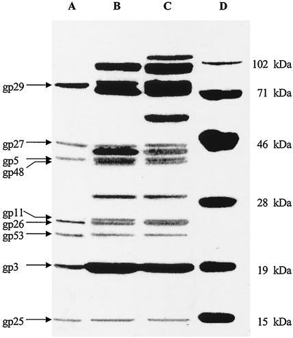
SDS-PAGE analysis of gene products from tube baseplates. The polyacrylamide concentration was 12.5%. Tube baseplates were treated with 6 M urea at 37°C for 15 min (lane A), with SDS buffer at 70°C for 3 min (lane B), or with SDS buffer at 90°C for 3 min (lane C). Lane D, protein molecular weight standards.
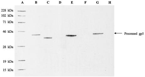
Recombinant gene 5-expressed protein products detected by Western blot analysis. After electrophoresis on SDS-12.5% PAGE, proteins were transferred to PVDF membranes. Lane A, Prestained protein molecular weight standards; lane B, gp5 separated from tube baseplates; lane C, sample from induced E. coli BL21(DE3) cells containing recombinant gene 5 with the codon for C-terminal Ser-360; lane D, sample from uninduced E. coli BL21(DE3) cells containing recombinant gene 5 with the codon for C-terminal Ser-360; lane E, sample from induced E. coli BL21(DE3) cells containing recombinant gene 5 with the codon for C-terminal Val-380; lane F, sample from uninduced E. coli BL21(DE3) cells containing recombinant gene 5 with the codon for C-terminal Val-380; lane G, sample from induced E. coli BL21(DE3) cells containing recombinant gene 5 with the codon for C-terminal Val-390; lane H, sample from uninduced E. coli BL21(DE3) cells containing recombinant gene 5 with the codon for C-terminal Val-390.
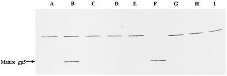
Western blot analysis of processed gp5 from lysates. After electrophoresis on SDS-8% PAGE, proteins were transferred to PVDF membranes. Lane A, 25am N67 lysate; lane B, 26am N131 lysate; lane C, 27am N120 lysate; lane D, 28am A452 lysate; lane E, 29am B7 lysate; lane F, tube baseplates; lane G, 5am B256 lysate; lane H, 48am NO22X lysate; lane I, 51am S29 lysate.

Western blot analysis of processed gp5 from lysates. After electrophoresis on SDS-8% PAGE, proteins were transferred to PVDF membranes. Lane A, prestained protein molecular mass standards (Bio-Rad Laboratories); lane B, 25am N67 lysate; lane C, 26am N131 lysate; lane D, 28am A452 lysate; lane E, 29am B7 lysate; lane F, 5am N135 lysate; lane G, 23am H11 lysate (tails); lane H, uninfected E. coli BE (sup0); lane I, 51am S29 lysate.
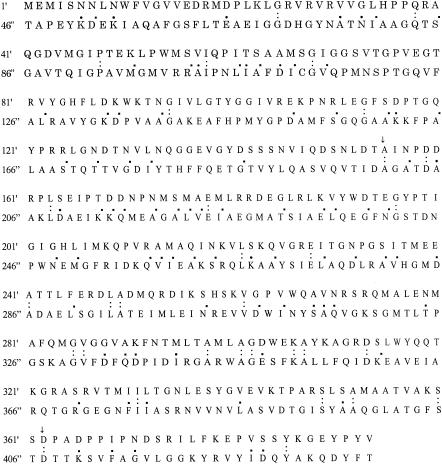
Similar regions of the amino acid sequence between gp5 (′) and gp23 ("). The ratio of identical amino acid pairs in whole gp5/gp23 (1′/46" to 390′/435") is 8.2%, much higher than 5.0% in two common proteins. When the number of conservative changes is included, the total similarity ratio of amino acid pairs between gp5 and gp23 reaches 30.0%. In the main region from 155′/200" to 362′/407" (208 amino acid pairs, the region indicated by arrows), the ratio of identical amino acid pairs in gp5 and gp23 is 11.5% and the total similarity ratio including conservative changes is 35.1%. Colons indicate identical amino acids. Periods indicate conservative changes.
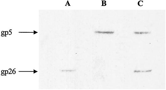
Western blot analysis of gp26. The tube baseplate proteins were transferred from SDS-12.5% PAGE gels to PVDF membranes. Lane A, gp26 band detected with antiserum against the C-terminal peptide of gp26; lane B, gp5 band detected with antiserum against gp5; lane C, gp26 and gp5 bands detected with a mixture of the two antisera.
Similar articles
-
The tail lysozyme complex of bacteriophage T4.Int J Biochem Cell Biol. 2003 Jan;35(1):16-21. doi: 10.1016/s1357-2725(02)00098-5. Int J Biochem Cell Biol. 2003. PMID: 12467643 Review.
-
Association and dissociation of the cell puncturing complex of bacteriophage T4 is controlled by both pH and temperature.Biochim Biophys Acta. 2006 Sep;1764(9):1487-92. doi: 10.1016/j.bbapap.2006.07.007. Epub 2006 Aug 2. Biochim Biophys Acta. 2006. PMID: 16956798
-
The C-terminal fragment of the precursor tail lysozyme of bacteriophage T4 stays as a structural component of the baseplate after cleavage.J Bacteriol. 1999 May;181(9):2739-44. doi: 10.1128/JB.181.9.2739-2744.1999. J Bacteriol. 1999. PMID: 10217762 Free PMC article.
-
Structure of the cell-puncturing device of bacteriophage T4.Nature. 2002 Jan 31;415(6871):553-7. doi: 10.1038/415553a. Nature. 2002. PMID: 11823865
-
The bacteriophage T4 DNA injection machine.Curr Opin Struct Biol. 2004 Apr;14(2):171-80. doi: 10.1016/j.sbi.2004.02.001. Curr Opin Struct Biol. 2004. PMID: 15093831 Review.
Cited by
-
Characterization of 29 newly isolated bacteriophages as a potential therapeutic agent against IMP-6-producing Klebsiella pneumoniae from clinical specimens.Microbiol Spectr. 2023 Sep 19;11(5):e0476122. doi: 10.1128/spectrum.04761-22. Online ahead of print. Microbiol Spectr. 2023. PMID: 37724861 Free PMC article.
-
Multifunctional roles of a bacteriophage phi 29 morphogenetic factor in assembly and infection.J Mol Biol. 2008 May 9;378(4):804-17. doi: 10.1016/j.jmb.2008.02.068. Epub 2008 Mar 7. J Mol Biol. 2008. PMID: 18394643 Free PMC article.
-
Broad-host-range Yersinia phage PY100: genome sequence, proteome analysis of virions, and DNA packaging strategy.J Bacteriol. 2008 Jan;190(1):332-42. doi: 10.1128/JB.01402-07. Epub 2007 Oct 26. J Bacteriol. 2008. PMID: 17965162 Free PMC article.
References
-
- Arisaka, F., S. Takeda, K. Funane, N. Nishijima, and S. Ishii. 1990. Structural studies of the contractile tail sheath protein of bacteriophage T4. 2. Structural analysis of the tail sheath protein, Gp18, by limited proteolysis, immunoblotting, and immunoelectron microscopy. Biochemistry 29:5057-5062. - PubMed
-
- Ben-Bassat, A., and K. Bauer. 1987. Amino-terminal processing of proteins. Nature 369:315.
-
- Black, L. W., M. K. Showe, and A. C. Steven. 1994. Morphogenesis of the T4 head, p. 218-258. In J. D. Karam (ed.), Molecular biology of bacteriophage T4. ASM Press, Washington, D.C.
-
- Coombs, D. H., and F. Arisaka. 1994. T4 tail structure and function, p. 259-281. In J. D. Karam (ed.), Molecular biology of bacteriophage T4. ASM Press, Washington, D.C.
MeSH terms
Substances
LinkOut - more resources
Full Text Sources
Miscellaneous
