. 2004 Sep 1;24(35):7754-61.
doi: 10.1523/JNEUROSCI.2588-04.2004.

Olfactory receptor proteins in axonal processes of chemosensory neurons

Affiliations

Olfactory receptor proteins in axonal processes of chemosensory neurons

Joerg Strotmann et al. J Neurosci. .

Abstract

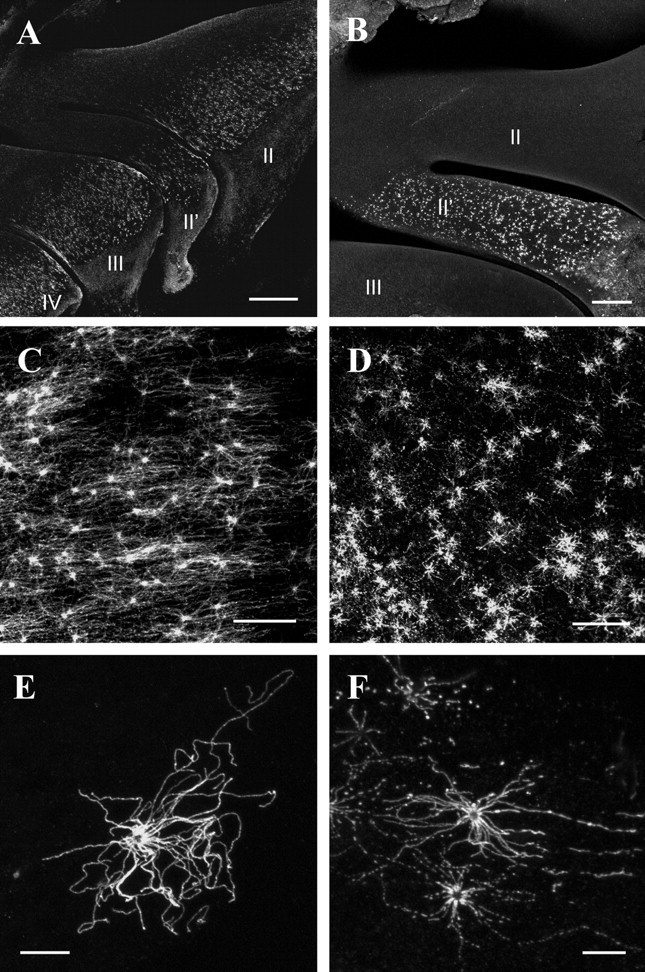
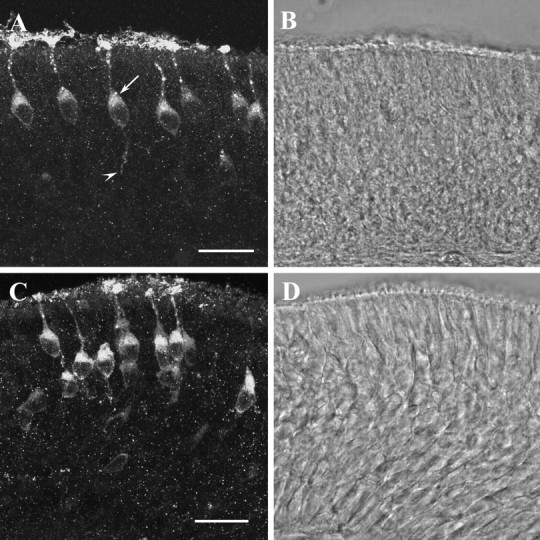
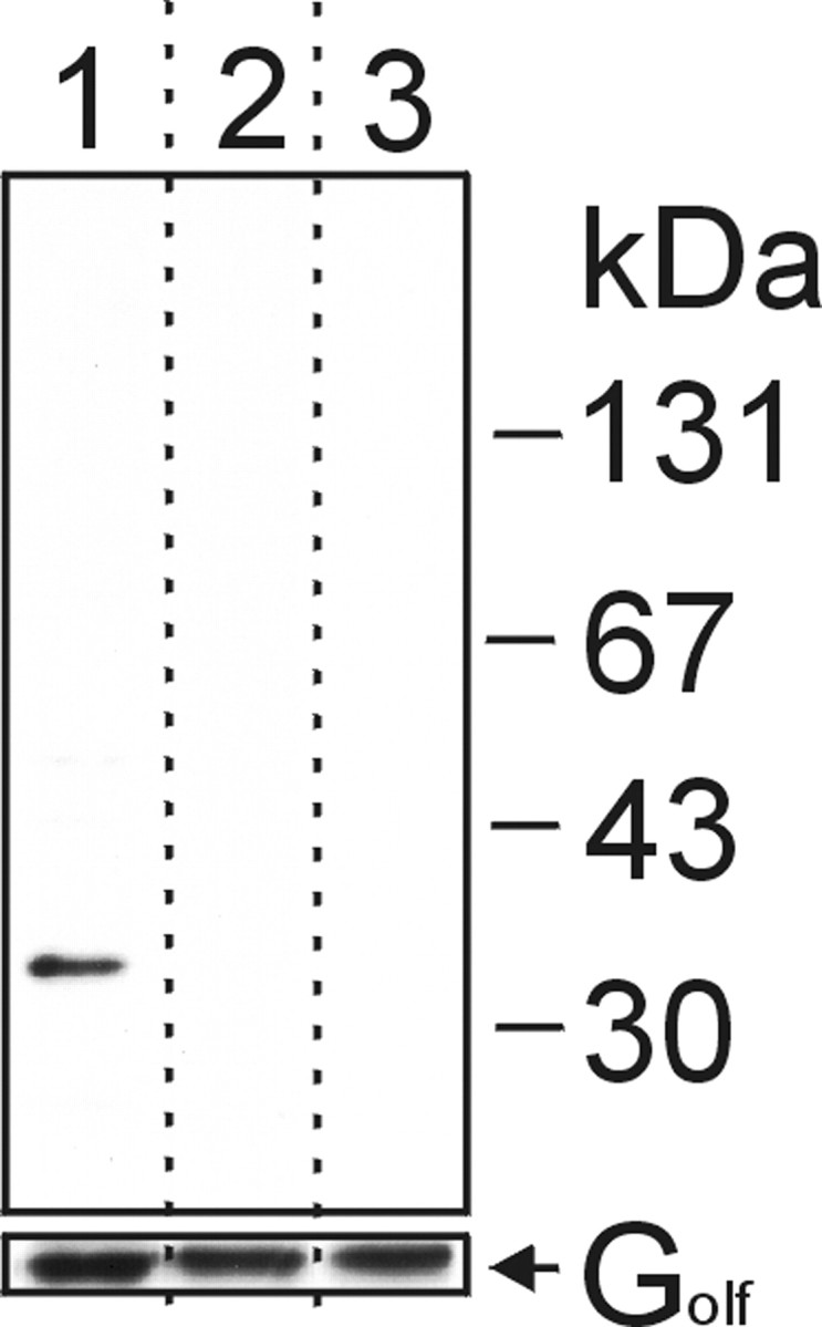
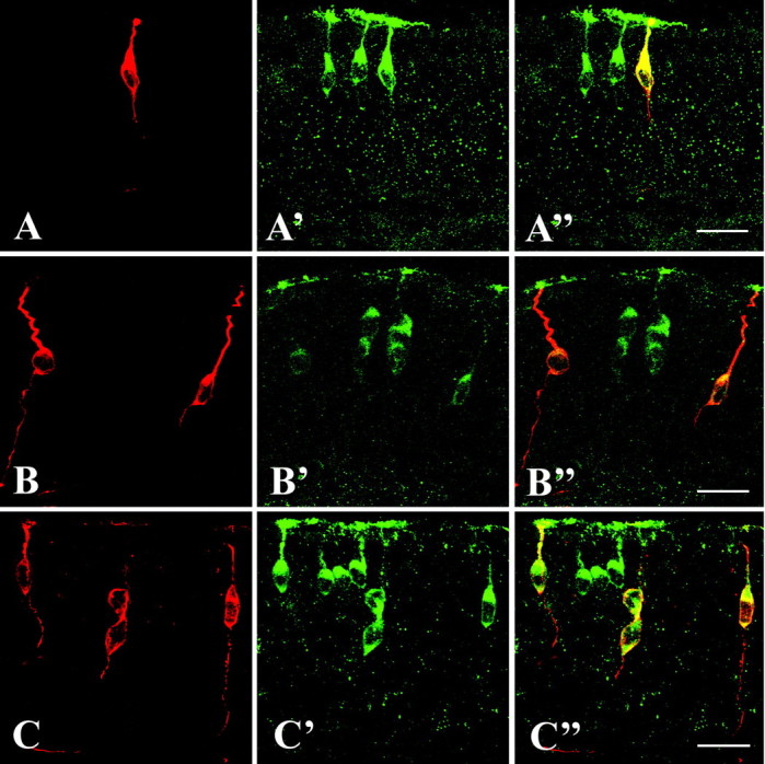
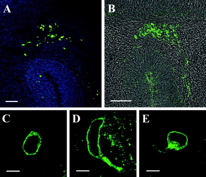
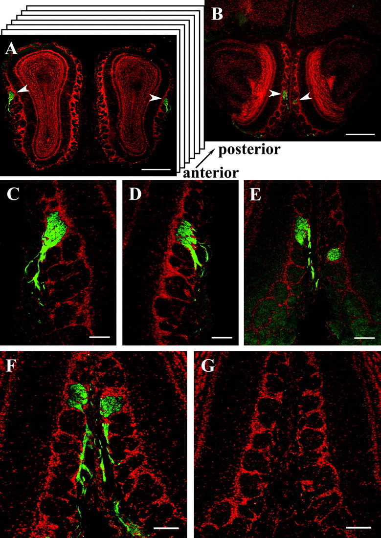
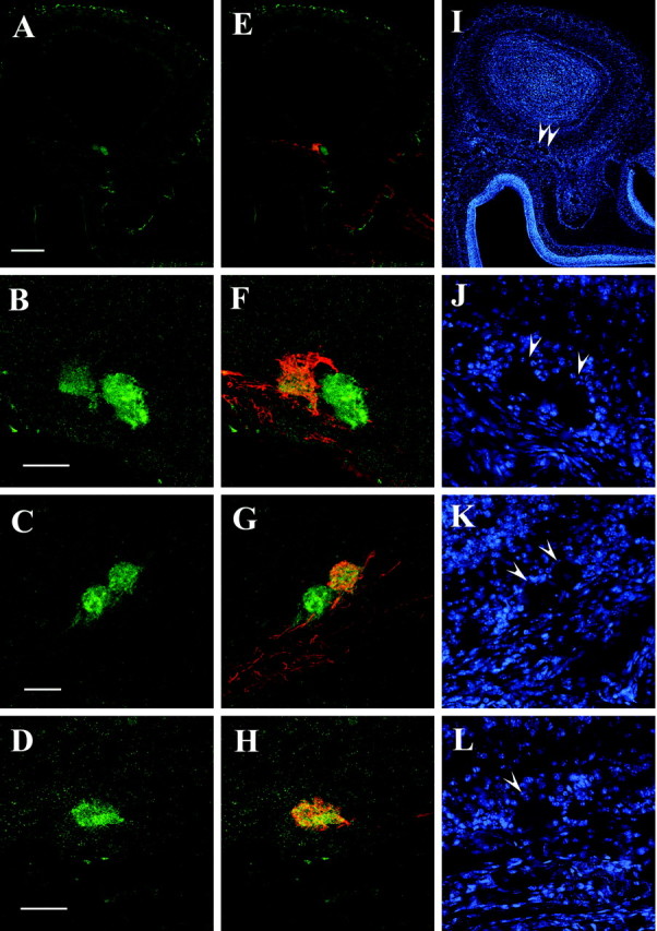
Olfactory receptors are supposed to act not only as molecular sensors for odorants but also as cell recognition molecules guiding the axons of olfactory neurons to their appropriate glomerulus in the olfactory bulb. This concept implies that olfactory receptor proteins are located in sensory cilia and in the axons. To approach this critical issue, antibodies were generated against two peptides, one derived from olfactory receptor mOR256-17, one derived from the "mOR37" subfamily. By means of immunohistochemistry and double-labeling studies using transgenic mouse lines as well as Western blot analyses, it was demonstrated that the newly generated antibodies specifically recognized the receptor proteins. To scrutinize the hypothesis that olfactory receptor proteins may also be present in the axonal processes and the nerve terminals, serial sections through the olfactory bulb were probed with the antibodies. Two glomeruli in each bulb were stained by anti-mOR256-17, one positioned in the medial, one in the lateral hemisphere. Fiber bundles approaching the glomeruli through the outer nerve layer also displayed intense immunofluorescence. A similar picture emerged for the antibody anti-mOR37, a small number of glomeruli in the ventral domain of the bulb was stained. On serial sections through the olfactory bulb of mOR37-transgenic mouse lines, double-labeling experiments demonstrated that distinct immunoreactive glomeruli corresponded to glomeruli that were targeted by neurons expressing a particular member of the mOR37 receptor subfamily. These data indicate that olfactory receptor (OR) proteins are indeed present in the axonal processes and nerve terminals of olfactory sensory neurons, thus supporting the notion that ORs may participate in the molecular processes underlying the fasciculation and targeting of olfactory axons.


Figures

Figure 1.
Figure 1.
A, B, View onto the whole-mount nasal turbinates probed with anti-mOR256-17 antibody (A) or anti-mOR37 antibody (B), respectively. II—IV, Endoturbinates. Scale bars: (in A) 500 μm; (in B) 200 μm. C, D, View onto the medial surface of endoturbinate II after incubation of anti-mOR256-17 antibody (C) or anti-mOR37 antibody (D). The knobs and cilia of distinct OSNs are strongly immunoreactive. Scale bars, 40 μm. E, F, High magnification of anti-mOR256-17-immunoreactive (E) or anti-mOR37-immunoreactive (F) OSN; the olfactory knob and cilia radiating in all directions are fluorescent. Scale bars, 10 μm.
Figure 2.
Figure 2.
A, Confocal image (16.8 μm stack) of a cross section through the olfactory epithelium of a mouse probed with the anti-mOR256-17 antibody; the strongest immunofluorescence is detectable in the knobs and cilia of individual OSNs. The dendrite and cell body of the cells are stained, as well. Most somata display a strong supranuclear staining (arrow). In some cases, the axonal process of individual cells (arrowhead) is visible extending into the deeper layer of the epithelium. Scale bar, 20 μm. B, Transmission channel picture of the cross section shown in A. C, Confocal image (14.6 μm) of a cross section through the olfactory epithelium probed with the anti-mOR37 antibody. A group of brightly immunoreactive OSNs is visible positioned in several layers of the epithelium; their cilia, knobs, and cell bodies are intensely labeled. Scale bar, 20 μm. D, Transmission channel picture of the cross section shown in C.
Figure 3.
Figure 3.
Western blot analysis using anti-mOR37 antibody. We loaded 10 μg of a ciliary membrane fraction prepared from mouse olfactory cilia on each lane of a polyacrylamide gel, subjected to SDS-PAGE, and transferred to a nitrocellulose membrane. After blotting, the lanes were separated (as indicated by dotted lines) and incubated with either anti-mOR37 (top panel, lane 1), anti-mOR37 preabsorbed to the corresponding antigenic blocking peptide (lane 2), or without primary antibody (lane 3). As a loading control, the same blot was reprobed with anti-Gs/olf antibody (bottom panel). Molecular weight markers are indicated.
Figure 4.
Figure 4.
A-A″, Cross section through the olfactory epithelium of an mOR37A-GFP transgenic mouse. Expression of mOR37A is visualized by intrinsic GFP fluorescence (A). Double labeling with the anti-mOR37 antibody visualized by an Alexa-568-labeled secondary antibody (A′). Overlay of both pictures (A″) shows that all mOR37A-expressing cells are immunoreactive for the anti-mOR37 antibody. Scale bar, 20 μm. B, B″, Cross section through the olfactory epithelium of an mOR37B-lacZ transgenic mouse probed with anti-β-galactosidase and Alexa-568-labeled secondary antibody to visualize mOR37B expression (B), and simultaneously with the anti-mOR37 antibody visualized by an Alexa-488-labeled secondary antibody (B′). Overlay of both pictures (B″) shows that all mOR37B-expressing cells visualized by lacZ expression are immunoreactive for the anti-mOR37 antibody. Scale bar, 20 μm. C-C″, Cross section through the olfactory epithelium of an mOR37C-lacZ transgenic mouse probed with anti-β-galactosidase and Alexa-568-labeled secondary antibody (C), and simultaneously with anti-mOR37 antibody visualized by an Alexa-488-labeled secondary antibody (C′). Overlay of both pictures (C″) shows that all mOR37C-expressing cells visualized by lacZ expression are immunoreactive for the anti-mOR37 antibody. Scale bar, 20 μm.
Figure 5.
Figure 5.
A, Cross section through the head of an E12.25 mouse embryo probed with an mOR256-17-specific, biotin-labeled antisense riboprobe. A group of extraepithelial receptor-expressing cells is located in the cribriform mesenchyme between the developing olfactory epithelium and the presumptive olfactory bulb. The section was counterstained with DAPI. Scale bar, 50 μm. B, Cross section through the head of an E12.25 mouse embryo probed with the anti-mOR256-17 antibody. A group of immunoreactive cells located in the same position as those cells detected by in situ hybridization (A) is visible. The immunofluorescence picture is overlayed with the transmission channel image. Scale bar, 50 μm. C, A 1 μm confocal section through an extraepithelial cell stained with the anti-mOR256-17 antibody. The cell exhibits a typical ring-shaped fluorescence. Scale bar, 5 μm. D, E, Confocal images (1 μm) of representative extraepithelial cells immunoreactive to anti-mOR256-17; both cells display a short process being immunopositive. Scale bars, 5 μm.
Figure 6.
Figure 6.
A, Cross section through the olfactory bulbs of a C57/BL6 wild-type mouse probed with the anti-mOR256-17 antibody. A strongly immunoreactive glomerulus is visualized in both bulbs in the dorsolateral domain (arrowheads). Counterstaining was performed with propidium iodide. Scale bar, 500 μm. B, Cross section through the bulbs shown in A from a more posterior region. A second pair of immunoreactive glomeruli (arrowheads) is visible in the medial region of both bulbs. Scale bar, 500 μm. C, Higher magnification of the glomerulus from the left bulb in A showing immunoreactive fibers in the outer nerve layer of the bulb entering the glomerulus. Scale bar, 100 μm. D, Higher magnification of the glomerulus from the right bulb in A showing immunoreactive fibers entering the glomerulus from the outer nerve layer. Scale bar, 100 μm. E, High-power photomicrograph of the pair of medial glomeruli located in the posterior region of the bulb. Immunoreactive fibers are only visible in the outer nerve layer of the left bulb; because of a slight skewing of the bulbs, the medial glomerulus in the right bulb is slightly displaced, and no fibers are visible on this section. Scale bar, 100 μm. F, Cross section with the pair of medial anti-mOR256-17-immunoreactive glomeruli and incoming fibers located in the posterior region of the bulbs. Scale bar, 100 μm. G, Adjacent cross section to F probed with peptide-preabsorbed anti-mOR265-17 antibody; no immunoreactivity is detectable. Scale bar, 100 μm.
Figure 7.
Figure 7.
A, Cross section through the olfactory bulb of an mOR37C-lacZ transgenic mouse probed with the anti-mOR37 antibody. Immunoreactivity is detectable in a small spot located the ventral region of the bulb. Scale bar, 200 μm. B, High magnification of the ventral domain of the bulb shown in A, visualizing the anti-mOR37-immunoreactive structures; two distinct glomeruli are stained located next to each other. Scale bar, 50 μm. C, High magnification of the ventral domain of the bulb from an mOR37B-lacZ transgenic mouse, visualizing the anti-mOR37-immunoreactive structures; two distinct glomeruli that are located next to each other are stained on this section. Scalebar, 50 μm. D, High magnification of the ventral domain of the bulb from an mOR37A-GFP transgenic mouse, visualizing the anti-mOR37 immunoreactive structure; one glomerulus is stained on this section. The glomerulus is visualized by an Alexa-568 conjugated antibody, which is presented in green color. Scale bar, 50 μm. E, Overlay of the picture shown in A with anti-β-galactosidase immunostaining demonstrating the position of axon terminals from OSNs expressing mOR37C; immunofluorescence is detectable in the ventral domain of the bulb, colocalized with the anti-mOR37 immunoreactivity. F, Overlay of the picture shown in B with anti-β-galactosidase immunostaining for mOR37C-lacZ; one of the two glomeruli on this section immunoreactive for anti-mOR37 contains fibers from mOR37C-expressing OSNs. G, Overlay of the picture shown in C with anti-β-galactosidase immunostaining for mOR37B-lacZ; one of the two glomeruli on this section immunoreactive for anti-mOR37 contains fibers from mOR37B-expressing OSNs. H, Overlay of the picture shown in D with in trinsic GFP fluorescence of mOR37A-GFP containing fibers; this glomerulus immunoreactive for anti-mOR37A also contains fibers from mOR37A-expressing OSNs. I, DAPI counterstaining of the section shown in A and E. Arrowheads indicate the positions of immunoreactive glomeruli. J, DAPI counterstaining of the section shown in B and F. Arrowheads indicate the positions of immunoreactive glomeruli. K, DAPI counterstaining of the section shown in C and G. Arrowheads indicate the positions of immunoreactive glomeruli. L, DAPI counterstaining of the section shown in D and H. Arrowhead indicates the position of the immunoreactive glomerulus.

References

    1. Anholt RRH, Aebi U, Snyder SH (1986) A partially purified preparation of isolated chemosensory cilia from the olfactory epithelium of the bullfrog, Rana catesbeiana J Neurosci 6: 1962-1969. - PMC - PubMed
    1. Barnea G, O'Donnell S, Mancia F, Sun X, Nemes A, Mendelson M, Axel R (2004) Odorant receptors on axon termini in the brain. Science 304: 1468. - PubMed
    1. Bozza TC, Mombaerts P (2001) Olfactory coding: revealing intrinsic representations of odors. Curr Biol 11: R687-R690. - PubMed
    1. Bozza T, Feinstein P, Zheng C, Mombaerts P (2002) Odorant receptor expression defines functional units in the mouse olfactory system. J Neurosci 22: 3033-3043. - PMC - PubMed
    1. Bradford MM (1976) A rapid and sensitive method for the quantitation of microquantites of protein utilizing the principle of protein-dye binding. Anal Biochem 65: 248-254. - PubMed

Publication types

MeSH terms