. 2004 Nov;15(11):4807-17.
doi: 10.1091/mbc.e04-05-0431. Epub 2004 Sep 1.

Regulation of cell motility by tyrosine phosphorylated villin

Affiliations

Regulation of cell motility by tyrosine phosphorylated villin

Alok Tomar et al. Mol Biol Cell. 2004 Nov.

Abstract

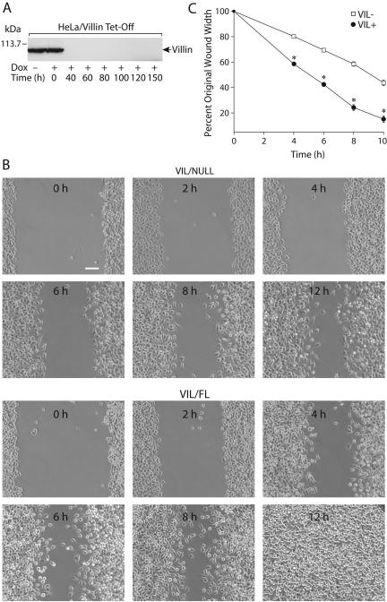
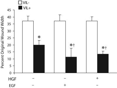
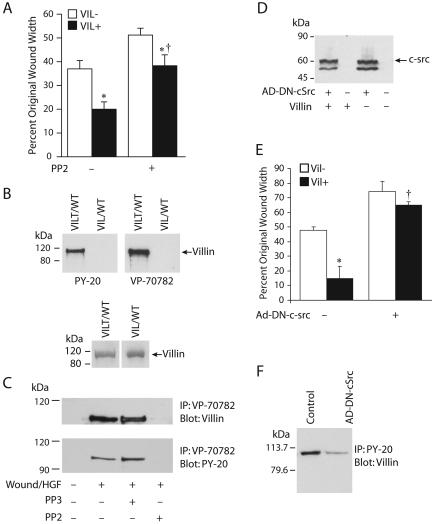
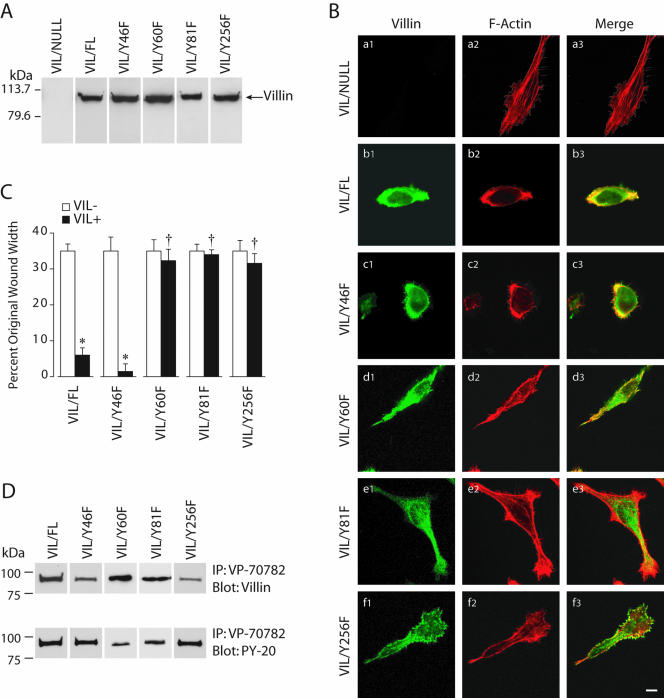
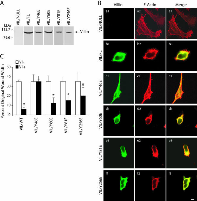
Temporal and spatial regulation of the actin cytoskeleton is vital for cell migration. Here, we show that an epithelial cell actin-binding protein, villin, plays a crucial role in this process. Overexpression of villin in doxycyline-regulated HeLa cells enhanced cell migration. Villin-induced cell migration was modestly augmented by growth factors. In contrast, tyrosine phosphorylation of villin and villin-induced cell migration was significantly inhibited by the src kinase inhibitor 4-amino-5-(4-chlorophenyl)-7-(t-butyl)pyrazolo[3,4-d]pyrimidine (PP2) as well as by overexpression of a dominant negative mutant of c-src. These data suggest that phosphorylation of villin by c-src is involved in the actin cytoskeleton remodeling necessary for cell migration. We have previously shown that villin is tyrosine phosphorylated at four major sites. To further investigate the role of tyrosine phosphorylated villin in cell migration, we used phosphorylation site mutants (tyrosine to phenylalanine or tyrosine to glutamic acid) in HeLa cells. We determined that tyrosine phosphorylation at residues 60, 81, and 256 of human villin played an essential role in cell migration as well as in the reorganization of the actin cytoskeleton. Collectively, these studies define how biophysical events such as cell migration are actuated by biochemical signaling pathways involving tyrosine phosphorylation of actin binding proteins, in this case villin.


Figures

Figure 1.
Figure 1.
HeLa Tet-Off cells expressing wild-type villin display significantly enhanced migration. (A) HeLa Tet-Off cells were stably transfected with full-length human villin cDNA. Cells cultured in the absence of doxycycline express villin (VIL/FL), whereas cells cultured in the presence of doxycycline (1.0 μg/ml) do not express villin (VIL/NULL). Expression of villin protein was followed 0–150 h after the addition of doxycycline. Western analysis was done using monoclonal villin antibodies. Data are representative of three experiments with similar results. (B) Equal numbers of wild-type (VIL/FL) and villin null (VIL/NULL) cells were cultured to confluence. Cells were denuded with a pipette tip and migration of the remaining cells into the wound is shown 0–12 h after the wound was introduced under low-serum condition (1.0% fetal bovine serum). Wound repair was measured as a percentage of initial wound area. Unlike villin-null cells, cells expressing villin seal the wound completely 12 h postwounding. Bar, 40 μm. (C) HeLa cells expressing villin migrate faster than villin-null cells at all time points. Migration distance was determined by taking two independent measurements from each well in a total of 24 wells. The error bars are the measured SEM and the asterisk denotes statistically significant values (p < 0.05, n = 24).
Figure 2.
Figure 2.
Villin-induced cell migration is enhanced by HGF and EGF. VIL/FL cells were treated without or with HGF (10 ng/ml) or EGF (10 nM) for 30 min and cell migration followed over a period of 6 h. Wound repair is expressed as a percentage of the initial wound area after 6 h. VIL (-) refers to each clone cultured in the presence of doxycycline, whereas VIL (+) refers to the same clone cultured in the absence of doxycycline. The error bars are the measured SEM, and the asterisk (*) and cross (†) denote statistically significant values [p < 0.05, n = 24, compared with VIL (-) cells] and (p < 0.05, n = 24, compared with untreated cells) respectively.
Figure 3.
Figure 3.
Villin-induced cell migration is regulated by c-src kinase. (A) The src kinase inhibitor PP2 inhibits villin-induced cell migration. VIL/FL cells were treated without or with the src-kinase–specific inhibitor PP2 (10 nM), and cell migration was followed over a period of 6 h. Wound repair is expressed as a percentage of the initial wound area after 6 h. VIL (-) refers to each clone cultured in the presence of doxycycline, whereas VIL (+) refers to the same clone cultured in the absence of doxycycline. The error bars are the measured SEM, and the asterisk (*) and cross (†) denotes statistically significant values compared with VIL (-) cells and untreated cells (p < 0.05, n = 24) respectively. (B) Characterization of phospho-villin antibody. Recombinant phosphorylated (VILT/WT) and nonphosphorylated (VIL/WT) villin proteins were used to determine the specificity of the phospho-villin antibody. The top panels show that tyrosine phosphorylated villin is immunodetected by the phospho-villin antibody VP-70782 (at 1:3000 dilution) as well as a commercially available anti-phosphotyrosine antibody, PY-20 (at 1:1000 dilution). The bottom panel is a control gel showing phosphorylated (VILT/WT) and nonphosphorylated (VIL/WT) villin proteins separated by SDS-PAGE and stained with GelCode Blue. Data are representative of three experiments with similar results. (C) HeLa cells cultured in the presence or absence of doxycycline were wounded (20 wounds/dish) and allowed to migrate in the presence of HGF (10 ng/ml) with or without the src kinase inhibitor PP2 (10 nM) or its inactive analog PP3 (10 nM). Tyrosine phosphorylated villin was immunoprecipitated using the phospho-villin antibody (VP-70782; 1 μg), and the Western blot was probed with villin mAb or phosphotyrosine antibody (PY-20). Data are representative of three experiments with similar results. (D) Expression of dominant negative c-src in VIL/NULL and VIL/FL cells. This is a Western blot with c-src monoclonal antibodies. (E) Dominant negative c-src inhibits villin-induced cell migration. VIL/FL and VIL/NULL cells were infected with recombinant adenovirus [Ad-DN-c-src (dominant negative c-src)] for 4 h at a multiplicity of infection of 100. Virus-containing media were then removed and cells were cultured for an additional 18 h to allow for expression of transgenic proteins. This was followed by measurement of cell migration. The error bars are the measured SEM and the asterisk (*) and cross (†) denote statistically significant values [p < 0.05, n = 24, compared with VIL (-) cells and (p < 0.05, n = 24, compared with untreated cells], respectively. (F) Dominant negative c-src inhibits the tyrosine phosphorylation of villin. Tyrosine phosphorylated villin was immunoprecipitated from VIL/FL cells infected with or without Ad-DN-c-src by using anti-phosphotyrosine antibody (clone PY-20), and Western analysis was done using a villin mAb. This is a representative of three experiments with similar results.
Figure 4.
Figure 4.
Tyrosine phosphorylation of villin is required for villin-induced increase in cell migration. (A) HeLa cells were stably transfected with wild-type (VIL/FL) and phosphorylation site mutants of villin, namely, Y46F, Y60F, Y81F, and Y256F. This figure shows representative clones of each villin construct transfected in HeLa cells. Data are representative of six experiments with similar results. (B) Villin expression results in reorganization of the actin cytoskeleton. HeLa cells transfected with wild-type (VIL/FL), and mutant villin proteins were analyzed by confocal microscopy. Double staining of villin (a1–f1) and F-actin (a2–f2) was performed using villin monoclonal antibodies (1:100) and FITC-conjugated anti-mouse IgG (1:200) and Alexa-Phalloidin 568 (1 μg/ml), respectively. Composite images of villin (green) and F-actin staining (red) are shown. Merged images (a3–f3) show colocalization of villin and F-actin. Wild-type villin and VIL/Y46F colocalize with F-actin at the cell periphery. In contrast, phosphorylation mutants of villin VIL/Y60F, VIL/Y81F, and VIL/Y256F show intracellular distribution of villin and F-actin with minimal colocalization of villin and F-actin at the cell surface. Bars, 3 μm. (C) HeLa cells expressing equal amounts of wild-type and phosphorylation site mutants (Y to F) of villin were used in wound-healing experiments. Wounds ∼800 μm in width were made using a pipette tip, and migration of cells into the wound was followed between 0 and 10 h postwounding. Wound repair is expressed as a percentage of the original wound width after 10 h. The error bars are the measured SEM, and the asterisk (*) and cross (†) denote statistically significant values [p < 0.05, n = 24, compared with VIL (-) cells and p < 0.05, n = 24, compared with untreated cells], respectively. VIL (-) refers to each clone cultured in the presence of doxycycline, whereas VIL (+) refers to the same clone cultured in the absence of doxycycline. (D) Tyrosine phosphorylation of wild-type and mutant villin proteins. Cell extracts from VIL/FL and the mutant villin cell lines VIL/Y46F, VIL/Y60F, VIL/Y81F, and VIL/Y256F were immunoprecipitated with phospho-villin antibody (VP-70782) and Western analysis done with villin mAb or phospho-tyrosine antibody (PY-20). This is not a quantitative Western blot. The Western blot is representative of three other experiments with similar results.
Figure 5.
Figure 5.
Phosphorylation of villin at Y-60, Y-81, and Y-256 is required for villin-induced cell migration. (A) HeLa cells were stably transfected with wild-type (VIL/FL) and phosphorylation site mutants of villin, namely, Y46E, Y60E, Y81E, and Y256E. This figure shows representative clones of each villin construct transfected in HeLa cells. Data are representative of six experiments with similar results. (B) Villin expression results in reorganization of the actin cytoskeleton. HeLa cells transfected with wild-type (VIL/FL) and mutant villin proteins were analyzed by confocal microscopy. Double staining of villin (a1–f1) and F-actin (a2–f2) was performed using villin monoclonal antibodies (1:100) and FITC conjugated anti-mouse IgG (1:200) and Alexa-Phalloidin 568 (1 μg/ml), respectively. Composite images of villin (green) and F-actin staining (red) are shown. Merged images show colocalization of villin and F-actin (a3–f3). Wild-type villin and VIL/Y60E, VIL/Y81E, and VIL/Y256E colocalize with F-actin at the cell periphery. In contrast, phosphorylation mutants of villin VIL/Y46E shows intracellular distribution of villin and F-actin with minimal colocalization of villin and F-actin at the cell surface. Bars, 3 μm. (C) HeLa cells expressing equal amounts of wild-type and phosphorylation site mutants (Y to E) of villin were used in wound-healing experiments. Wounds ∼800 μm in width were made using a pipette tip, and migration of cells into the wound was followed between 0 and 10 h postwounding. Wound repair is expressed as a percentage of the original wound width at 10 h. The error bars are the measured SEM, and the asterisk (*) and cross (†) denote statistically significant values [p < 0.05, n = 24, compared with VIL (-) cells and p < 0.05, n = 24, compared with untreated cells], respectively. VIL (-) refers to each clone cultured in the presence of doxycycline, whereas VIL (+) refers to the same clone cultured in the absence of doxycycline.

Similar articles

Cited by

References

    1. Aizawa, H., Sutoh, K., and Yahara, I. (1996). Overexpression of cofilin stimulates bundling of actin filaments, membrane ruffling, and cell movement in Dictyostelium. J. Cell Biol. 132, 335-344. - PMC - PubMed
    1. Anderson, R.D., Haskell, R.E., Xia, H., Roessler, B.J., and Davidson, B.L. (2000). A simple method for the rapid generation of recombinant adenovirus vectors. Gene Ther. 7, 1034-1038. - PubMed
    1. Arora, P.D., and McCulloch, C.A. (1996). Dependence of fibroblast migration on actin severing activity of gelsolin. J. Biol. Chem. 271, 20516-20523. - PubMed
    1. Arpin, M., Pringault, E., Finidori, J., Garcia, A., Jeltsch, J.M., Vandekerckhove, J., and Louvard, D. (1988). Sequence of human villin: a large duplicated domain homologous with other actin-severing proteins and a unique small carboxy-terminal domain related to villin specificity. J. Cell Biol. 107, 1759-1766. - PMC - PubMed
    1. Athman, R., Louvard, D., and Robine, S. (2003). Villin enhances hepatocyte growth factor-induced actin cytoskeleton remodeling in epithelial cells. Mol. Biol. Cell 14, 4641-4653. - PMC - PubMed

Publication types

MeSH terms