

Contribution of bone marrow-derived cells to skin: collagen deposition and wound repair
- PMID: 15342945
- PMCID: PMC1388268
- DOI: 10.1634/stemcells.22-5-812
Contribution of bone marrow-derived cells to skin: collagen deposition and wound repair
Abstract
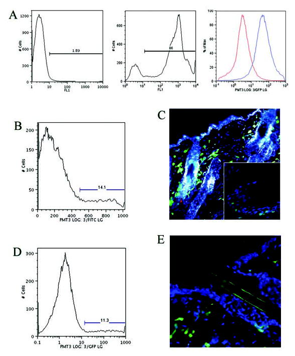
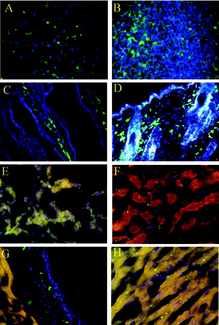
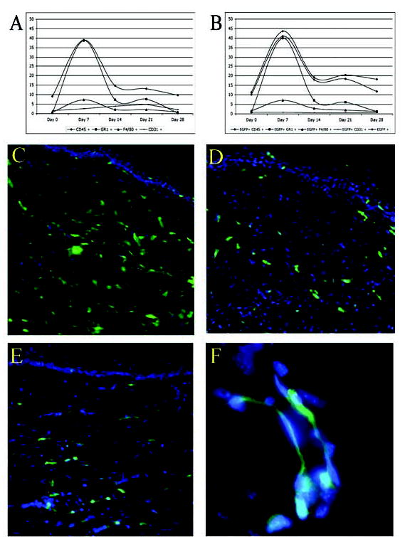
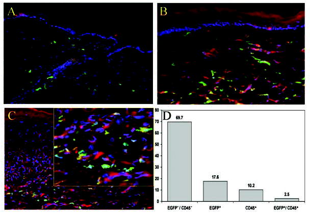
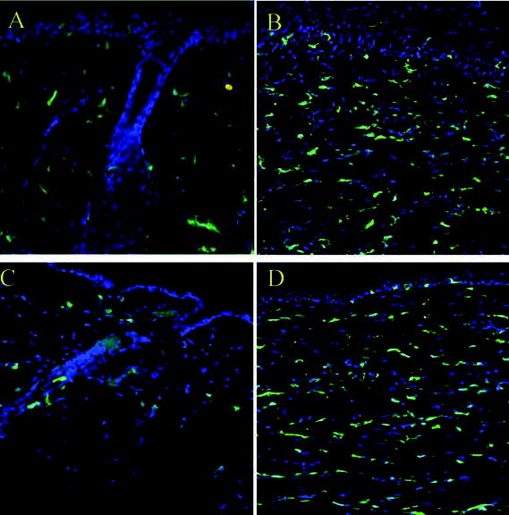
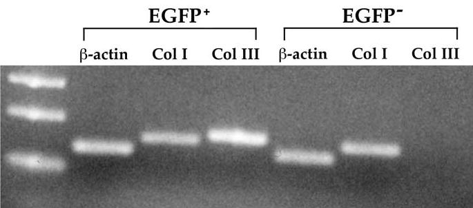
The bone marrow provides inflammatory cells and endothelial progenitor cells to healing cutaneous wounds. To further explore the bone marrow contribution to skin and healing wounds, we used a chimeric mouse model in which the bone marrow from enhanced green fluorescent protein (EGFP) transgenic mice is transplanted into normal C57BL mice. We found that normal skin is a target organ for bone marrow-derived cells from both the hematopoietic and the mesenchymal stem cell pool. We present evidence that the bone marrow contribution to normal skin and the healing cutaneous wound is substantially greater than the previously recognized CD45+ subpopulation, where 15%-20% of the spindle-shaped dermal fibroblasts were bone marrow-derived (EGFP+). Furthermore, the bone marrow-derived cells were able to contract a collagen matrix and transcribe both collagen types I and III, whereas the skin-resident cells transcribed only collagen type I. Whereas endothelial progenitor cells were found early during the wound repair process, bone marrow-derived endothelial cells were not seen after epithelialization was complete. Our data show that wound healing involves local cutaneous cells for reconstituting the epidermis but distant bone marrow-derived cells and the adjacent uninjured dermal mesenchymal cells for reconstituting the dermal fibroblast population.
Figures
Similar articles
-
Fibroblasts/myofibroblasts that participate in cutaneous wound healing are not derived from circulating progenitor cells.J Cell Physiol. 2010 Mar;222(3):703-12. doi: 10.1002/jcp.21997. J Cell Physiol. 2010. PMID: 20020505
-
Differential contribution of dermal resident and bone marrow-derived cells to collagen production during wound healing and fibrogenesis in mice.J Invest Dermatol. 2011 Feb;131(2):529-36. doi: 10.1038/jid.2010.314. Epub 2010 Oct 21. J Invest Dermatol. 2011. PMID: 20962852
-
Bone marrow-derived cells in the healing burn wound--more than just inflammation.Burns. 2009 May;35(3):356-64. doi: 10.1016/j.burns.2008.07.011. Epub 2008 Oct 25. Burns. 2009. PMID: 18952376
-
Use of mesenchymal stem cells for cutaneous repair and skin substitute elaboration.Pathol Biol (Paris). 2014 Apr;62(2):108-17. doi: 10.1016/j.patbio.2014.01.002. Epub 2014 Mar 21. Pathol Biol (Paris). 2014. PMID: 24661975 Review.
-
Concise review: bone marrow-derived stem/progenitor cells in cutaneous repair and regeneration.Stem Cells. 2010 May;28(5):905-15. doi: 10.1002/stem.420. Stem Cells. 2010. PMID: 20474078 Free PMC article. Review.
Cited by
-
Accelerated wound healing in a diabetic rat model using decellularized dermal matrix and human umbilical cord perivascular cells.Acta Biomater. 2016 Nov;45:234-246. doi: 10.1016/j.actbio.2016.08.053. Epub 2016 Aug 31. Acta Biomater. 2016. PMID: 27591919 Free PMC article.
-
Genesis of the myofibroblast in lung injury and fibrosis.Proc Am Thorac Soc. 2012 Jul;9(3):148-52. doi: 10.1513/pats.201201-011AW. Proc Am Thorac Soc. 2012. PMID: 22802289 Free PMC article. Review.
-
Inhibitory effect of anti-aminopeptidase N/CD13 antibodies on fibroblast migration.Mol Cell Biochem. 2010 Oct;343(1-2):191-9. doi: 10.1007/s11010-010-0513-7. Epub 2010 Jun 30. Mol Cell Biochem. 2010. PMID: 20589526 Free PMC article.
-
A Systematic Review of the Evidence of Hematopoietic Stem Cell Differentiation to Fibroblasts.Biomedicines. 2022 Nov 28;10(12):3063. doi: 10.3390/biomedicines10123063. Biomedicines. 2022. PMID: 36551819 Free PMC article. Review.
-
Phenotypic Modulation of Adipose-Derived Stem Cells and Fibroblasts Treated with Povidone-Iodine and Chlorhexidine in Mono and Coculture Models.Biomedicines. 2023 Jun 29;11(7):1855. doi: 10.3390/biomedicines11071855. Biomedicines. 2023. PMID: 37509495 Free PMC article.
References
-
- Singer AJ, Clark RA. Cutaneous wound healing. N Engl J Med. 1999;341:738–746. - PubMed
-
- Prockop DJ. Marrow stromal cells as stem cells for non-hematopoietic tissues. Science. 1997;276:71–74. - PubMed
-
- Krause DS, Theise ND, Collector MI, et al. Multi-organ, multi-lineage engraftment by a single bone marrow–derived stem cell. Cell. 2001;105:369–377. - PubMed
-
- Lagasse E, Connors H, Al-Dhalimy M, et al. Purified hematopoietic stem cells can differentiate into hepatocytes in vivo. Nat Med. 2000;6:1229–1234. - PubMed
-
- Orlic D, Kajstura J, Chimenti S, et al. Bone marrow cells regenerate infarcted myocardium. Nature. 2001;410:701–705. - PubMed
Publication types
MeSH terms
Substances
Grants and funding
LinkOut - more resources
Full Text Sources
Other Literature Sources
Medical
Research Materials
Miscellaneous
