Review
. 2004 Sep;4(9):699-710.
doi: 10.1038/nri1439.

NALT- versus Peyer's-patch-mediated mucosal immunity

Affiliations
Review

NALT- versus Peyer's-patch-mediated mucosal immunity

Hiroshi Kiyono et al. Nat Rev Immunol. 2004 Sep.

Abstract

Recent studies indicate that the mechanism of nasopharynx-associated lymphoid tissue (NALT) organogenesis is different from that of other lymphoid tissues. NALT has an important role in the induction of mucosal immune responses, including the generation of T helper 1 and T helper 2 cells, and IgA-committed B cells. Moreover, intranasal immunization can lead to the induction of antigen-specific protective immunity in both the mucosal and systemic immune compartments. Therefore, a greater understanding of the differences between NALT and other organized lymphoid tissues, such as Peyer's patches, should facilitate the development of nasal vaccines.


Conflict of interest statement

The authors declare no competing financial interests.

Figures
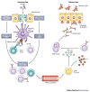
Figure 1
Figure 1. The common mucosal immune system.
Luminal antigens are transported to the nasopharynx-associated lymphoid tissue (NALT) and Peyer's patches through microfold (M) cells that are present in the epithelium overlying NALT and Peyer's-patch follicles. Dendritic cells process and present antigens to T cells in these lymphoid tissues. CD4+ T cells that are stimulated by dendritic cells then preferentially induce IgA-committed B-cell development in the germinal centre of the lymphoid follicle. After IgA class switching and affinity maturation, B cells rapidly migrate from NALT and Peyer's patches to the regional cervical lymph nodes and mesenteric lymph nodes respectively, through the efferent lymphatics. Finally, antigen-specific CD4+ T cells and IgA+ B cells migrate to effector sites (such as the nasal passage and intestinal lamina propria) through the thoracic duct and blood circulation. IgA+ B cells and plasmablasts then differentiate into IgA-producing plasma cells in the presence of cytokines (such as interleukin-5 (IL-5) and IL-6) that are produced by T helper 2 (TH2) cells, and they subsequently produce dimeric (or polymeric) forms of IgA. These dimeric forms of IgA then become secretory IgA by binding to polymeric Ig receptors (which become the secretory component in the process of secretory IgA formation) that are displayed on the monolayer of epithelial cells lining the mucosa. Secretory IgA is then released into the nasal passage and intestinal tract. TCR, T-cell receptor.
Figure 2
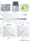
Figure 2. Chronological differences between NALT- and Peyer's-patch tissue genesis.
a | Nasal tissue from newborn mice (day 0) is characterized by an absence of peripheral-node addressin (PNAD)-expressing high endothelial venules (HEVs). The NASOPHARYNX-ASSOCIATED LYMPHOID-TISSUE (NALT) ANLAGEN from one-week-old mice shows a small accumulation of lymphoid cells around a single PNAD-expressing HEV in the nasal tissue. In eight-week-old mice, NALT contains numerous PNAD-expressing HEVs. This figure is reproduced with permission from Ref. © Elsevier (2002). b | The formation of NALT therefore starts after birth, whereas the development of Peyer's patches is initiated during embryogenesis. c | These kinetic differences in the initiation of tissue genesis of NALT and Peyer's patches are also supported by the appearance and frequency of CD3CD4+CD45+ inducer cells in nasal and intestinal tissues. The inducer cells accumulate postnatally at the site of NALT formation, whereas high numbers of these cells are observed in Peyer's patches during the gestational period. E, embryonic day.
Figure 3
Figure 3. Comparison of the organogenesis programme of NALT and Peyer's patches.
CD3CD4+CD45+ cells are considered to be the common inducers of secondary lymphoid tissue. ID2 (inhibitor of DNA binding 2) is indispensable for the induction and differentiation of these inducer cells from their fetal-liver precursors (which have the phenotype CD3CD4CD45+). a | For Peyer's patches, after activation through the interleukin-7 receptor (IL-7R) or TRANCE (tumour-necrosis-factor-related activation-induced cytokine), these CD3CD4+CD45+ cells express the lymphotoxin-α1β2 (LT-α1β2) heterotrimer, which then binds to the LT-β receptor (LT-βR) displayed on stromal cells and induces signal transduction through NIK (nuclear factor-κB (NF-κB)-inducing kinase). In turn, NIK promotes the expression of adhesion molecules and/or chemokines. These homing molecules trigger the accumulation of lymphoid cells at the site of Peyer's patches. So, the IL-7R- and LT-βR-mediated signals are essential for the tissue genesis of Peyer's patches. b | The development of CD3CD4+CD45+ cells in nasopharynx-associated lymphoid tissue (NALT) also requires ID2; however, the initiation of NALT organogenesis is independent of signalling that involves the IL-7R, LT-α1β2–LT-βR interactions and NIK. CCL, CC-chemokine ligand; CXCL, CXC-chemokine ligand; ICAM1, intercellular adhesion molecule 1; IKK-α, inhibitor of NF-κB (IκB) kinase-α; ROR-γ, retinoic-acid-receptor-related orphan receptor-γ; VCAM1, vascular cell-adhesion molecule 1.
Figure 4
Figure 4. Model for the induction of organogenesis of NALT and Peyer's patches by two subsets of CD3CD4+CD45+ inducer cells.
CD3CD4+CD45+ cells differentiate from fetal-liver progenitors. We propose that both ROR-γ (retinoic-acid-receptor-related orphan receptor-γ) and ID2 (inhibitor of DNA binding 2) are essential for the generation of interleukin-7 receptor (IL-7R)-expressing CD3CD4+CD45+ cells for the induction of Peyer's-patch organogenesis. By contrast, the generation of the IL-7RCD3CD4+CD45+ inducer cells that are involved in nasopharynx-associated lymphoid tissue (NALT) organogenesis is regulated by ID2 but not ROR-γ. However, this model remains to be tested experimentally, and other possibilities exist (see main text). NALT genesis in ID2-deficient mice can be initiated by the adoptive transfer of CD3CD4+CD45+ cells from wild-type mice, but the maturation of NALT formation is incomplete. This indicates that other cell populations, or the endogenous expression of ID2 at the site of NALT, might be required for the full maturation of NALT.

References

    1. Mestecky J, Blumberg R, Kiyono H, McGhee JR. Fundamental Immunology. 2003. pp. 965–1020.
    1. Yuki Y, Kiyono H. New generation of mucosal adjuvants for the induction of protective immunity. Rev. Med. Virol. 2003;13:293–310. doi: 10.1002/rmv.398. - DOI - PubMed
    1. Csencsits KL, Jutila MA, Pascual DW. Nasal-associated lymphoid tissue: phenotypic and functional evidence for the primary role of peripheral node addressin in naive lymphocyte adhesion to high endothelial venules in a mucosal site. J. Immunol. 1999;163:1382–1389. - PubMed
    1. Adachi S, Yoshida H, Kataoka H, Nishikawa S. Three distinctive steps in Peyer's patch formation of murine embryo. Int. Immunol. 1997;9:507–514. doi: 10.1093/intimm/9.4.507. - DOI - PubMed
    1. Fukuyama S, et al. Initiation of NALT organogenesis is independent of the IL-7R, LTβR, and NIK signaling pathways but requires the Id2 gene and CD3−CD4+CD45+ cells. Immunity. 2002;17:31–40. doi: 10.1016/S1074-7613(02)00339-4. - DOI - PubMed

Publication types