. 2004 Sep;114(5):679-88.
doi: 10.1172/JCI21075.

Anti-C1q autoantibodies deposit in glomeruli but are only pathogenic in combination with glomerular C1q-containing immune complexes

Affiliations

Anti-C1q autoantibodies deposit in glomeruli but are only pathogenic in combination with glomerular C1q-containing immune complexes

Leendert A Trouw et al. J Clin Invest. 2004 Sep.

Abstract

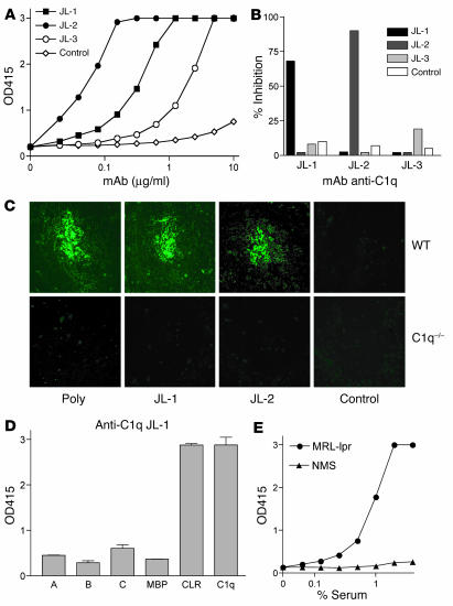
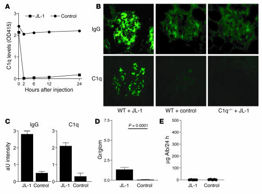
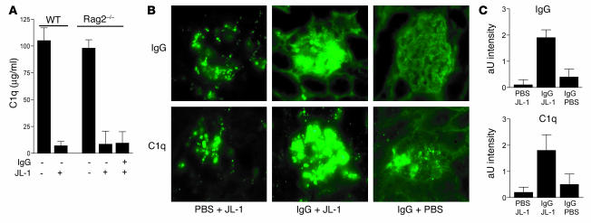
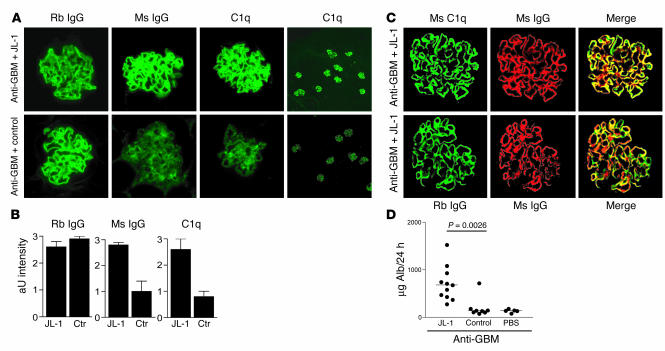
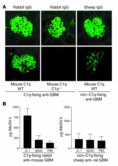
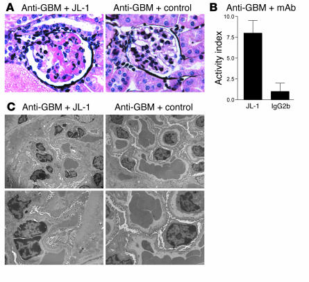
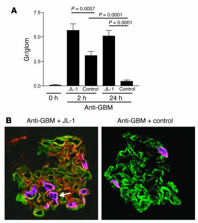
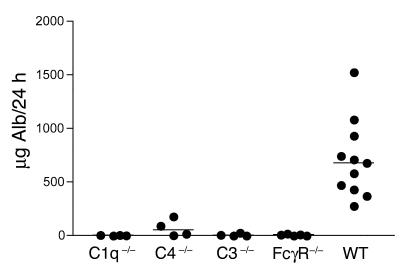
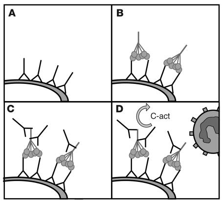
Anti-C1q autoantibodies are present in sera of patients with several autoimmune diseases, including systemic lupus erythematosus (SLE). Strikingly, in SLE the presence of anti-C1q is associated with the occurrence of nephritis. We have generated mouse anti-mouse C1q mAb's and used murine models to investigate whether anti-C1q autoantibodies actually contribute to renal pathology in glomerular immune complex disease. Administration of anti-C1q mAb JL-1, which recognizes the collagen-like region of C1q, resulted in glomerular deposition of C1q and anti-C1q autoantibodies and mild granulocyte influx, but no overt renal damage. However, combination of JL-1 with a subnephritogenic dose of C1q-fixing anti-glomerular basement membrane (anti-GBM) antibodies enhanced renal damage characterized by persistently increased levels of infiltrating granulocytes, major histological changes, and increased albuminuria. This was not observed when a non-C1q-fixing anti-GBM preparation was used. Experiments with different knockout mice showed that renal damage was dependent not only on glomerular C1q and complement activation but also on Fcgamma receptors. In conclusion, anti-C1q autoantibodies deposit in glomeruli together with C1q but induce overt renal disease only in the context of glomerular immune complex disease. This provides an explanation why anti-C1q antibodies are especially pathogenic in patients with SLE.


Figures

Figure 1
Figure 1
In vitro characterization of mouse anti–mouse C1q mAb. (A) Anti-C1q detection ELISA for anti-C1q mAb’s JL-1, JL-2, and JL-3 and control mAb IgG2b under 0.5 M NaCl buffer conditions. OD415, OD at 415 nm. (B) Epitope competition ELISA showing inhibition of binding of DIG-labeled anti-C1q mAb to mouse C1q by unlabeled anti-C1q mAb or control mAb IgG2b. (C) Immunohistochemistry of WT or C1q–/– mouse spleen stained with anti-C1q mAb or controls. Original magnification, ×250. Poly, polyclonal antibody. (D) C1q head domains of the human A, B, and C chains or the control maltose-binding protein (MBP), CLRs, and intact human C1q were coated, and mAb JL-1 binding was analyzed. (E) Anti-C1q tail ELISA using human CLRs and serum of autoimmune MRL-lpr mice (MRL-lpr) or nonautoimmune normal mouse serum (NMS) for binding.
Figure 2
Figure 2
Effects of administration of anti-C1q mAb to naive mice. (A) C1q sandwich ELISA for the detection of serum C1q levels following administration of anti-C1q mAb and control mAb.(B) Immunofluorescence analysis of mouse kidneys stained for the presence of mouse IgG and mouse C1q following administration of JL-1 or control mAb to WT or C1q–/– mice. Original magnification, ×400. (C) Quantification of immunofluorescence analysis of the deposition of IgG and C1q in WT mice. Data are expressed as arbitrary units (aU), as described in Methods. (D) Quantification of glomerular granulocyte influx. Depicted on the y axis are granulocytes per glomerular cross section (Gr/glom) at 24 hours after injection. (E) Assessment of albuminuria by ELISA. Alb, albumin.
Figure 3
Figure 3
Anti-C1q autoantibodies react with C1q in the glomerulus in a planted antigen–like fashion. (A) C1q sandwich ELISA for serum levels of C1q in naive WT mice, WT mice treated with JL-1, naive Rag2–/– mice, and Rag2–/– mice pretreated with PBS or with mouse IgG and then treated with JL-1. (B) Immunofluorescence analysis of renal sections of Rag2–/– mice, either naive or reconstituted for mouse IgG followed by injection of either JL-1 or PBS. Images show the absence of IgG and C1q in naive Rag2–/– mice and positivity for IgG and C1q, both mesangially and along the GBM, in IgG-reconstituted mice. The positivity for IgG and C1q is highly increased following administration of JL-1. Original magnification, ×400. (C) Quantification of immunofluorescence analysis of the glomerular deposition of IgG and C1q.
Figure 4
Figure 4
Effects of administration of anti-C1q mAb to mice pretreated with C1q-fixing anti-GBM antibodies. (A) Immunofluorescence of renal sections obtained from mice pretreated with rabbit (Rb) anti-GBM antibodies combined with either mAb JL-1 or IgG2b control mAb stained for the presence of mouse (Ms) IgG, mouse C1q, or rabbit IgG. Images show linear, GBM-like deposition of anti-GBM antibodies, linear fixation of C1q and anti-C1q in the JL-1–coinjected mice, and only mild mesangial positivity for C1q and anti-C1q in control-coinjected mice. Original magnification, ×400. Right panels: Low-power magnifications of renal sections of mice injected with anti-GBM in combination with either JL-1 or control and stained for C1q. Original magnification, ×100. (B) Quantification of immunofluorescence analysis of the glomerular deposition of rabbit IgG, mouse IgG, and C1q. Ctr, control. (C) Confocal analysis of kidney sections of mice injected with rabbit anti-GBM and JL-1. Representative pictures are shown for the colocalization (yellow) of mouse C1q (green) and mouse IgG (red) and the colocalization (yellow) of rabbit IgG (green) and mouse IgG (red). They indicate that rabbit IgG, mouse C1q, and mouse IgG do colocalize in these mice, in a linear, GBM-like pattern. (D) Albuminuria of mice injected with anti-GBM antibodies followed by JL-1, control mAb, or PBS.
Figure 5
Figure 5
The disease-enhancing effect of JL-1 is dependent on glomerular C1q. (A) Immunofluorescence analysis of anti-GBM antibody deposition and C1q deposition for the C1q-fixing anti-GBM preparation in WT and C1q–/– mice and the non–C1q-fixing anti-GBM preparation in WT mice. The images show the linear IgG fixation for both anti-GBM preparations, but only linear fixation of C1q for the rabbit anti-GBM preparation. Only some mild mesangial C1q positivity can be observed in the mice given sheep anti-GBM. Original magnification, ×400. (B) Groups of WT mice were injected with either the C1q-fixing or the non–C1q-fixing anti-GBM preparation and coinjected with JL-1, control, or PBS, and albuminuria was determined.
Figure 6
Figure 6
Histological changes induced by JL-1 at both the light microscopic and the electron microscopic levels. (A) Histological analysis of Silver-stained renal sections of mice injected with rabbit anti–mouse GBM and coinjected with either mAb JL-1 or IgG2b control mAb, obtained at 24 hours after injection. For JL-1–coinjected mice, images show pronounced inflammatory cell influx, focal capillary tuft occlusion by microthrombi, necrotizing lesions, nuclear debris, and wireloop-like lesions. Control-coinjected mice only display marginal inflammatory cell influx. Original magnification, ×400. (B) Quantification of histological changes using the activity index as described in Methods. (C) Electron microscopic analysis of glomerular lesions of mice injected with rabbit anti–mouse GBM and coinjected with either anti-C1q mAb JL-1 or control mAb IgG2b. At higher magnification, we observed several wireloop-like lesions in the JL-1–coinjected mice, whereas the IgG2b-coinjected mice did not display any abnormalities. Original magnifications, ×2,000 (left) and ×4,000 (right).
Figure 7
Figure 7
Quantification of glomerular granulocyte influx. (A) Mice were injected with anti-GBM antibodies in combination with either JL-1 or control mAb. Granulocytes per glomerular cross section were scored either at 2 hours or at 24 hours after injection. (B) Confocal analysis of sections stained for mouse C1q (green), mouse IgG (red), and mouse granulocytes (purple). The pictures are merged and show, in yellow, colocalization of green and red, and, in white, colocalization of green and purple. The anti-GBM antibodies induced linear fixation of C1q in both groups, but only in the JL-1–coinjected mice is there colocalization between C1q and IgG and a pronounced influx of granulocytes. Original magnification, ×400. The white arrow indicates the white colocalization between C1q and granulocytes.
Figure 8
Figure 8
Anti-GBM and JL-1 in various genetically deficient mice. Rabbit anti–mouse GBM in combination with JL-1 was administered to either WT C57BL/6 mice or C1q–/–, C4–/–, C3–/–, or FcγR–/– mice, and albuminuria was assessed.
Figure 9
Figure 9
Schematic representation of the pathogenic role of anti-C1q antibodies in immune complex–mediated renal disease. (A) Deposition of immune complexes on the GBM. (B) Fixation of C1q from the circulation by the immune complexes. (C) Binding of anti-C1q autoantibodies to C1q present on the immune complexes. (D) Activation of complement (C-Act) and attraction of inflammatory cells.

Comment in

Similar articles

Cited by

References

    1. Lipsky PE. Systemic lupus erythematosus: an autoimmune disease of B cell hyperactivity. Nat. Immunol. 2001;2:764–766. - PubMed
    1. Mok CC, Lau CS. Pathogenesis of systemic lupus erythematosus. J. Clin. Pathol. 2003;56:481–490. - PMC - PubMed
    1. Mills JA. Systemic lupus erythematosus. N. Engl. J. Med. 1994;330:1871–1879. - PubMed
    1. Tsao BP. The genetics of human systemic lupus erythematosus. Trends Immunol. 2003;24:595–602. - PubMed
    1. Walport MJ. Complement. First of two parts. N. Engl. J. Med. 2001;344:1058–1066. - PubMed

Publication types

MeSH terms