Meta-Analysis
. 2004;5(9):R66.
doi: 10.1186/gb-2004-5-9-r66. Epub 2004 Aug 12.

Discovery of estrogen receptor alpha target genes and response elements in breast tumor cells

Affiliations
Meta-Analysis

Discovery of estrogen receptor alpha target genes and response elements in breast tumor cells

Chin-Yo Lin et al. Genome Biol. 2004.

Abstract

Background: Estrogens and their receptors are important in human development, physiology and disease. In this study, we utilized an integrated genome-wide molecular and computational approach to characterize the interaction between the activated estrogen receptor (ER) and the regulatory elements of candidate target genes.

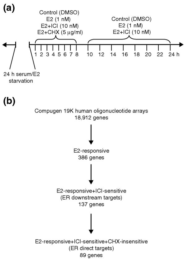
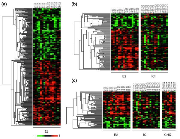
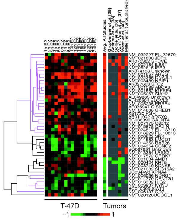
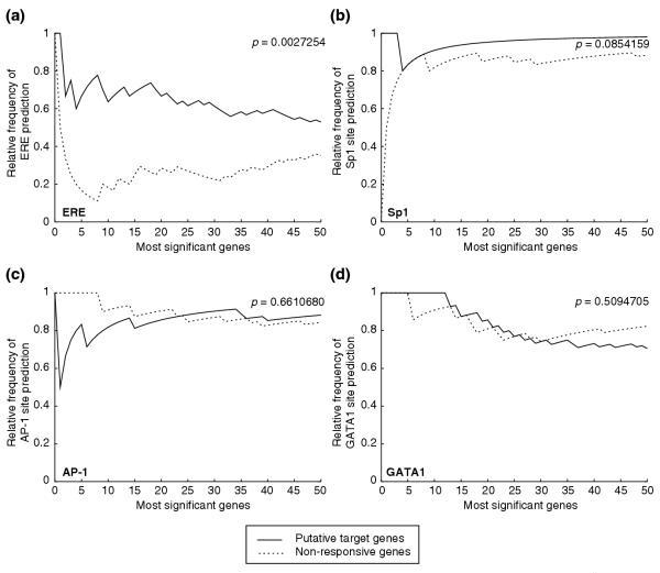
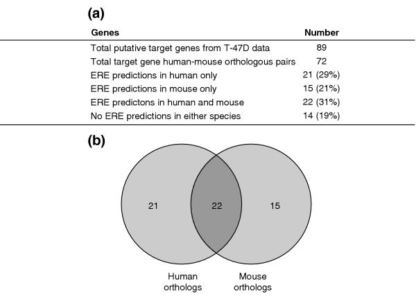
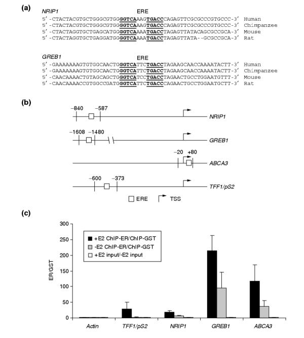
Results: Of around 19,000 genes surveyed in this study, we observed 137 ER-regulated genes in T-47D cells, of which only 89 were direct target genes. Meta-analysis of heterogeneous in vitro and in vivo datasets showed that the expression profiles in T-47D and MCF-7 cells are remarkably similar and overlap with genes differentially expressed between ER-positive and ER-negative tumors. Computational analysis revealed a significant enrichment of putative estrogen response elements (EREs) in the cis-regulatory regions of direct target genes. Chromatin immunoprecipitation confirmed ligand-dependent ER binding at the computationally predicted EREs in our highest ranked ER direct target genes, NRIP1, GREB1 and ABCA3. Wider examination of the cis-regulatory regions flanking the transcriptional start sites showed species conservation in mouse-human comparisons in only 6% of predicted EREs.

Conclusions: Only a small core set of human genes, validated across experimental systems and closely associated with ER status in breast tumors, appear to be sufficient to induce ER effects in breast cancer cells. That cis-regulatory regions of these core ER target genes are poorly conserved suggests that different evolutionary mechanisms are operative at transcriptional control elements than at coding regions. These results predict that certain biological effects of estrogen signaling will differ between mouse and human to a larger extent than previously thought.


Figures

Figure 1
Figure 1
Experimental design and microarray data selection of the time-course analysis of estrogen response in T-47D cells. (a) Cells were starved of serum and estrogen for 24 h before treatment with dimethysulfoxide (DMSO; carrier control), 17β-estradiol (E2), and E2 in combination with ICI 182,780 (ICI) and cycloheximide (CHX). Samples were taken at the 16 time points indicated. (b) Procedure for identifying direct ER targets. Data selection for estrogen-responsive genes was based on p-value cutoffs (p ≤ 0.052) and magnitude of response (at least 1.2-fold change in the same direction for three time points). ICI sensitivity and CHX insensitivity were also determined by statistical measures, p ≤ 0.058 and p ≥ 0.24 respectively. Cutoff values were informed by expression profiles of cathespsin D and progesterone receptor, both known to be regulated by ER.
Figure 2
Figure 2
Expression profiles of estrogen-responsive genes. (a) The 386 genes responsive to E2 were visualized by hierarchical clustering and the Eisen TreeView software. The columns represent time points arranged in chronological order, and each row represents the expression profile of a particular gene. By convention, upregulation is indicated by a red signal and downregulation by green. The magnitude of change is proportional to the brightness of the signal. Each panel represents a treatment condition as noted in the column headings. (b) ICI and (c) CHX treatments were included to identify the 137 ER-regulated genes and 89 primary response genes, respectively.
Figure 3
Figure 3
Comparative analysis of estrogen-responsive gene expression profiles in T-47D and MCF-7 breast cancer cell lines revealed similar responses. (a) Expression data of 103 genes that were responsive to E2 and sensitive to ICI treatment in T47-D cells and present in the MCF-7 dataset [31] revealed concordant responses to E2 in 64% (66/103) of the genes (highlighted in magenta). (b) If the MCF-7 data is selected for E2-responsive and tamoxifen (Tam)-sensitive genes (see Materials and methods), there is a 24-gene overlap between the two datasets and the responses to E2 and the anti-estrogens ICI or tamoxifen are highly concordant (magenta) at nearly 96% (23/24).
Figure 4
Figure 4
Estrogen-responsive genes identified in cell-line studies were also differentially expressed in ER+ breast tumors compared to ER- tumors. Estrogen-responsive gene-expression profiles are compared to the composite expression ratio for those genes in each of the six breast tumor studies surveyed (L.D.M., B.M.F. Mow, L.A.V., and E.T.L., unpublished work and [36-40]). Differentially expressed genes were defined by p < 0.01 between ER+ and ER- tumor samples. Genes that responded similarly in ER+ tumors and in vitro following E2 treatment are highlighted in magenta.
Figure 5
Figure 5
ERE-like sequences are enriched in the extended promoter regions of putative target gene. (a) ERE predictions were made using a previously published model and optimized sensitivity setting. Prediction models were tested on the extended promoter regions (-3,000 to +500, both strands) from the most significant putative ER target and non-responsive genes, ordered by statistical significance. The y-axis represents the relative frequency of binding-site predictions as determined by the number of genes with predicted sites divided by the total number of genes. The number of most significant genes queried is indicated on the x-axis. Frequency of ERE predictions in putative target genes is significantly greater (p = 0.0027) than the similarly ranked non-responsive genes. Binding-site predictions were also carried out using position weight matrices describing sites for (b) Sp1, (c) AP-1 and (d) GATA1. Both Sp1 and AP-1 are known to be involved in regulating ER binding in certain target genes. GATA1 sites were included as negative controls. There is no significant enrichment of these sites in the putative target genes (see p-values in figure).
Figure 6
Figure 6
Comparison of human and mouse orthologs. (a) Statistics from the comparative analysis of predicted EREs in human and mouse orthologous putative target gene pairs. (b) Venn diagram showing that out of the 72 orthologous pairs extracted for analysis, only 22 pairs have ERE predictions made in both the human and mouse sequences.
Figure 7
Figure 7
ER binds promoter regions encoding both conserved and non-conserved predicted response elements in an estrogen-dependent manner. (a) EREs (underlined) found upstream of NRIP1 and GREB1 coding regions are conserved in human, chimpanzee, mouse, and rat genomes. (b) PCR primers flanking the predicted conserved (NRIP1 and GREB1) and non-conserved (ABCA3 and TFF1/pS2) EREs were designed to detect ER binding following ChIP assays. The relative positions of the primers and ERE, relative to the TSS, are indicated. (c) Interactions between ER and predicted EREs were enhanced by estrogen treatment. MCF-7 cells were either mock-treated with the carrier dimethyl sulfoxide (-E2, gray bars) or treated with estradiol (+E2, black bars), followed by ChIP experiments. Black and gray bars indicate the enrichment of the binding site in anti-ER ChIP experiments over anti-GST ChIP experiments. Enrichment of all EREs was observed in hormone-treated cells whereas the mock-treated cells displayed less or very little enrichment. There was no enrichment of actin exon 3 control region or any of the input controls (open bars).

References

    1. Korach KS, Emmen JM, Walker VR, Hewitt SC, Yates M, Hall JM, Swope DL, Harrell JC, Couse JF. Update on animal models developed for analyses of estrogen receptor biological activity. J Steroid Biochem Mol Biol. 2003;86:387–391. doi: 10.1016/S0960-0760(03)00348-0. - DOI - PubMed
    1. Ali S, Coombes RC. Estrogen receptor alpha in human breast cancer: occurrence and significance. J Mammary Gland Biol Neoplasia. 2000;5:271–281. doi: 10.1023/A:1009594727358. - DOI - PubMed
    1. Persson I. Estrogens in the causation of breast, endometrial and ovarian cancers - evidence and hypotheses from epidemiological findings. J Steroid Biochem Mol Biol. 2000;74:357–364. doi: 10.1016/S0960-0760(00)00113-8. - DOI - PubMed
    1. Nilsson S, Gustafsson JÅ. Estrogen receptor action. Crit Rev Eukaryot Gene Expr. 2002;12:237–257. - PubMed
    1. Klinge CM. Estrogen receptor interaction with estrogen response elements. Nucleic Acids Res. 2001;29:2905–2919. doi: 10.1093/nar/29.14.2905. - DOI - PMC - PubMed

MeSH terms