Comparative Study
. 2004 Sep;87(3):1752-66.
doi: 10.1529/biophysj.104.044552.

Oleic and docosahexaenoic acid differentially phase separate from lipid raft molecules: a comparative NMR, DSC, AFM, and detergent extraction study

Affiliations
Comparative Study

Oleic and docosahexaenoic acid differentially phase separate from lipid raft molecules: a comparative NMR, DSC, AFM, and detergent extraction study

Saame Raza Shaikh et al. Biophys J. 2004 Sep.

Abstract

We have previously suggested that the omega-3 polyunsaturated fatty acid, docosahexaenoic acid (DHA) may in part function by enhancing membrane lipid phase separation into lipid rafts. Here we further tested for differences in the molecular interactions of an oleic (OA) versus DHA-containing phospholipid with sphingomyelin (SM) and cholesterol (CHOL) utilizing (2)H NMR spectroscopy, differential scanning calorimetry, atomic force microscopy, and detergent extractions in model bilayer membranes. (2)H NMR and DSC (differential scanning calorimetry) established the phase behavior of the OA-containing 1-[(2)H(31)]palmitoyl-2-oleoyl-sn-glycero-3-phosphoethanolamine (16:0-18:1PE-d(31))/SM (1:1) and the DHA-containing 1-[(2)H(31)]palmitoyl-2-docosahexaenoyl-sn-glycero-3-phosphoethanolamine (16:0-22:6PE-d(31))/SM (1:1) in the absence and presence of equimolar CHOL. CHOL was observed to affect the OA-containing phosphatidylethanolamine (PE) more than the DHA-containing PE, as exemplified by >2 x greater increase in order measured for the perdeuterated palmitic chain in 16:0-18:1PE-d(31)/SM (1:1) compared to 16:0-22:6PE-d(31)/SM (1:1) bilayers in the liquid crystalline phase. Atomic force microscopy (AFM) experiments showed less lateral phase separation between 16:0-18:1PE-rich and SM/CHOL-rich raft domains in 16:0-18:1PE/SM/CHOL (1:1:1) bilayers than was observed when 16:0-22:6PE replaced 16:0-18:1PE. Differences in the molecular interaction of 16:0-18:1PE and 16:0-22:6PE with SM/CHOL were also found using biochemical detergent extractions. In the presence of equimolar SM/CHOL, 16:0-18:1PE showed decreased solubilization in comparison to 16:0-22:6PE, indicating greater phase separation with the DHA-PE. Detergent experiments were also conducted with cardiomyocytes fed radiolabeled OA or DHA. Although both OA and DHA were found to be largely detergent solubilized, the amount of OA that was found to be associated with raft-rich detergent-resistant membranes exceeded DHA by almost a factor of 2. We conclude that the OA-PE phase separates from rafts far less than DHA-PE, which may have implications for cellular signaling.


Figures
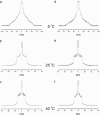
FIGURE 1
FIGURE 1
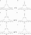
2H NMR spectra for 50 wt % aqueous dispersions in 50 mM Tris buffer (pH 7.4) of 16:0-18:1PE-d31/SM (1:1) and 16:0-22:6PE-d31/SM (1:1). Spectra for 16:0-18:1PE-d31/SM (1:1) were recorded at (a) 5, (b) 25, and (c) 40°C. Spectra for 16:0-22:6PE-d31/SM (1:1) were also recorded at (d) 5, (e) 25, and (f) 40°C.
FIGURE 2
FIGURE 2
2H NMR spectra for 50 wt % aqueous dispersions in 50 mM Tris buffer (pH 7.4) of 16:0-18:1PE-d31/SM/CHOL (1:1:1) and 16:0-22:6PE-d31/SM/CHOL (1:1:1). Spectra for 16:0-18:1PE-d31/SM/CHOL (1:1:1) were recorded at (a) 5, (b) 25, and (c) 40°C. Spectra for 16:0-22:6PE-d31/SM/CHOL were also recorded at (d) 5, (e) 25, and (f) 40°C.
FIGURE 3
FIGURE 3
(a and b) Variation in the first moment M1 (Eq. 1) as a function of temperature for bilayers of (a) 16:0-18:1PE-d31/SM (1:1) and 16:0-18:1PE-d31/SM/CHOL (1:1:1), and (b) 16:0-22:6PE-d31/SM (1:1) and 16:0-22:6PE-d31/SM/CHOL (1:1:1). M1 is plotted logarithmically and values have a ±2% error. The transition temperature Tm, designated by X, is the midpoint of the sharp drop in moment that accompanies the chain-melting transition. (c and d) Corresponding DSC cooling scans for (c) 16:0-18:1PE/SM (1:1) and 16:0-18:1PE/SM/CHOL (1:1:1), and (d) 16:0-22:6PE/SM (1:1) and 16:0-22:6PE/SM/CHOL (1:1:1). The scans with cholesterol are slightly offset along the excess heat capacity axis to aid observation. The inset is a high-resolution deconvolution of the DSC cooling scan for 16:0-18:1PE/SM (1:1). The dotted and dashed lines represent the 16:0-18:1PE-rich and SM-rich phases, respectively.
FIGURE 4
FIGURE 4
FFT depaked spectra for (a) 16:0-18:1PE-d31/SM (1:1); (b) 16:0-18:1PE-d31/SM/CHOL (1:1:1); (c) 16:0-22:6PE-d31/SM (1:1); and (d) 16:0-22:6PE-d31/SM/CHOL (1:1:1) at 40°C.
FIGURE 5
FIGURE 5
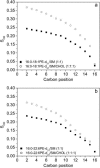
Smoothed order parameter profiles generated from depaked spectra at 40°C for bilayers of (a) 16:0-18:1PE-d31/SM (1:1) and 16:0-18:1PE-d31/SM/CHOL (1:1:1), and (b) 16:0-22:6PE-d31/SM (1:1) and 16:0-22:6PE-d31/SM/CHOL (1:1:1).
FIGURE 6
FIGURE 6
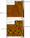
Representative AFM images at 23°C of 16:0-18:1PE/SM/CHOL (1:1:1) (top) and 16:0-22:6PE/SM/CHOL (1:1:1) (bottom) supported bilayers with a z scale of 20 nm (color bar). The insets present the section profile corresponding to the white line on the image.
FIGURE 7
FIGURE 7
Percentage (mean + SD) of SM, CHOL, and PE found in the DSM fraction of Triton X-100 treated aqueous multilamellar dispersions of 16:0-18:1PE/SM/CHOL (1:1:1) (no pattern) or 16:0-22:6PE/SM/CHOL (1:1:1) (dense pattern) at (a) 4 and (b) 40°C. Each value is relative to the total amount of the specific lipid in both detergent-soluble and detergent-insoluble fractions. The values represent a minimum of four samples from three separate experiments. Supernatant lipids were recovered by HPTLC and quantified via charring (Materials and Methods).
FIGURE 8
FIGURE 8
Percentage (mean + SD) of (a) GM1, (b) [3H]OA, and (c) [14C]DHA in sucrose-gradient fractions of neonatal cardiomyocytes. Cells were incubated at 4°C in the presence of 1% Triton X-100, homogenized, and placed on a 5–40% sucrose gradient. After ultracentrifugation, fractions were collected in 1-mL increments from the top to bottom of the centrifuge tubes and analyzed (Materials and Methods). Values represent a minimum of three separate experiments.
FIGURE 9
FIGURE 9
Cartoon depiction of DHA-induced alteration of plasma membrane lipid domain structure. The outer membrane leaflet is shown since it is here that the SM- and cholesterol-rich lo (raft) domains exist in a background of nonraft bulk lipids. Upon the addition of a DHA-rich phospholipid, cholesterol is excluded from the nonraft (ld) domains into the raft domains, increasing raft size and stability. In the example presented here, the inactive protein, X, initially found in the raft domain, is then recruited to the DHA-rich, nonraft domain where it undergoes a conformational change, resulting in its activation.

References

    1. Ahmed, S. N., D. A. Brown, and E. London. 1997. On the origin of sphingolipid/cholesterol-rich detergent insoluble cell membranes: physiological concentrations of cholesterol and sphingolipid induce formation of a detergent-insoluble, liquid-ordered lipid phase in model membranes. Biochemistry. 36:10944–10953. - PubMed
    1. Aussenac, F., M. Tavares, and E. J. Dufourc. 2003. Cholesterol dynamics in membranes of raft composition: a molecular point of view from 2H and 31P solid-state NMR. Biochemistry. 42:1383–1390. - PubMed
    1. Brown, D. A., and E. London. 2000. Structure and function of sphingolipid and cholesterol-rich membrane rafts. J. Biol. Chem. 275:17221–17224. - PubMed
    1. Brzustowicz, M. R., V. Cherezov, M. Caffrey, W. Stillwell, and S. R. Wassall. 2002b. Molecular organization of cholesterol in polyunsaturated membranes: microdomain formation. Biophys. J. 82:285–298. - PMC - PubMed
    1. Brzustowicz, M. R., V. Cherezov, M. Zerouga, M. Caffrey, W. Stillwell, and S. R. Wassall. 2002a. Controlling membrane cholesterol content. A role for polyunsaturated (docosahexaenoate) phospholipids. Biochemistry. 41:12509–12519. - PubMed

Publication types

MeSH terms

LinkOut - more resources