. 2004 Sep;87(3):1836-47.
doi: 10.1529/biophysj.104.044123.

Organization of Ca2+ release units in excitable smooth muscle of the guinea-pig urinary bladder

Affiliations

Organization of Ca2+ release units in excitable smooth muscle of the guinea-pig urinary bladder

Edwin D Moore et al. Biophys J. 2004 Sep.

Erratum in

  • Biophys J. 2004 Oct;87(4):2914

Abstract

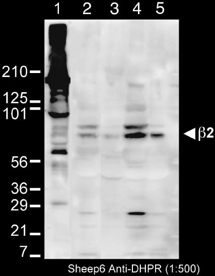
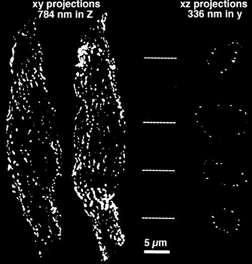
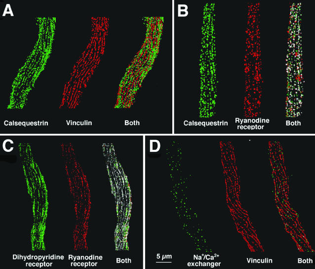
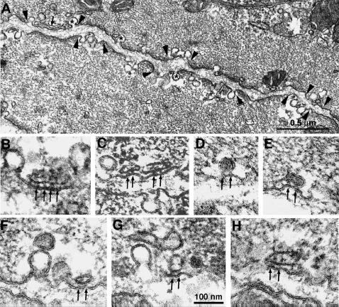
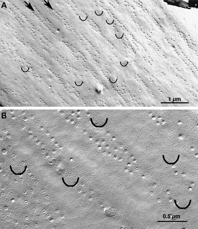
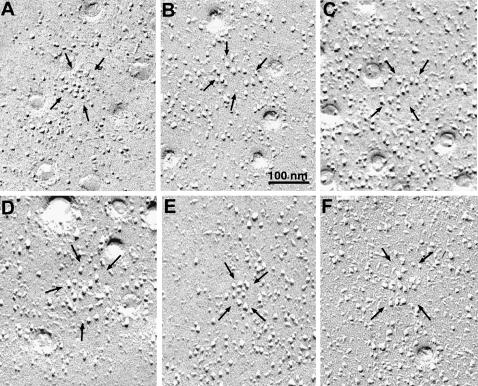
Ca(2+) release from internal stores (sarcoplasmic reticulum or SR) in smooth muscles is initiated either via pharmaco-mechanical coupling due to the action of an agonist and involving IP3 receptors, or via excitation-contraction coupling, mostly involving L-type calcium channels in the plasmalemma (DHPRs), and ryanodine receptors (RyRs), or Ca(2+) release channels of the SR. This work focuses attention on the structural basis for the coupling between DHPRs and RyRs in phasic smooth muscle cells of the guinea-pig urinary bladder. Immunolabeling shows that two proteins of the SR: calsequestrin and the RyR, and one protein the plasmalemma, the L-type channel or DHPR, are colocalized with each other within numerous, peripherally located sites located within the caveolar domains. Electron microscopy images from thin sections and freeze-fracture replicas identify feet in small peripherally located SR vesicles containing calsequestrin and distinctive large particles clustered within small membrane areas. Both feet and particle clusters are located within caveolar domains. Correspondence between the location of feet and particle clusters and of RyR- and DHPR-positive foci allows the conclusion that calsequestrin, RyRs, and L-type Ca(2+) channels are associated with peripheral couplings, or Ca(2+) release units, constituting the key machinery involved in excitation-contraction coupling. Structural analogies between smooth and cardiac muscle excitation-contraction coupling complexes suggest a common basic mechanism of action.


Figures

FIGURE 1
FIGURE 1
Western blot of a membrane preparation from rabbit skeletal muscle (1) and of several fractions from a crude preparation of membranes from the guinea pig urinary bladder (25). The sheep6 anti-DHPR (L-type Ca channel) serum that was used in the immunolabeling recognizes two bands at ∼100 and 60 kDa in the rabbit SR (Pragnell et al., 1991; Arikkath et al., 2003), corresponding to α1 and β-subunits and a band at ∼70 kDa in the smooth muscle crude membranes, which is about the size of the β2 subunit. The crude sheep antisera made against a rabbit skeletal muscle preparation combined with a rabbit anti-sheep secondary antibody results in a nonspecific reaction against high molecular weight components the rabbit membranes (lane 1). Other nonspecific bands (e.g., the ∼27 kDa band in lane 4, which may be the light chain of the antibody being picked up by the secondary are absent from the purified membrane fraction, lane 5). (Lane 1) Membrane fraction from rabbit skeletal muscle; (Lane 2) guinea pig urinary bladder homogenate; (Lane 3) 14,000 × g pellet; (Lane 4) 30,000 × g pellet; and (Lane 5) final membrane fraction.
FIGURE 2
FIGURE 2
Fluorescent images of a single enzymatically dissociated smooth muscle cell from the guinea pig urinary bladder immunolabeled with an antibody against vinculin. These images have been deconvolved and thresholded; no other image processing steps were performed. The two images on the left are xy projections in which the cells were bisected (along the xy plane) to prevent superposition of the front and back surfaces. The images on the right are xz projections of the same cells, but after having been rotated 90° round the x axis; the images are 336 nm deep in y and were acquired from the locations indicated by the dashed lines. The images show that vinculin is clearly located at the cell surface, where it is distributed in longitudinal stripes. The scale bar is 5 μm.
FIGURE 3
FIGURE 3
Double immunofluorescence images comparing the distribution of vinculin, calsequestrin, ryanodine receptor, L-type Ca channel (β-subunit) and the Na+/Ca2+ exchanger in single dissociated cells from the guinea-pig bladder. As in Fig. 2, one-half of the cell is displayed. For each of the image pairs, one image has been pseudocolored red and the other green. In the overlapped images, the voxels are colored white if they have identical three-dimensional coordinates for the two single images; otherwise, the voxel remain green or red. Since intensity information cannot be converted into number of protein copies in a given voxel, the intensity information has been removed from the data. Voxel that were above threshold are either green or red, and overlapped voxels are pure white. (A) Calsequestrin (green) and vinculin (red) are located at foci that occupy separate longitudinal stripes of membrane. (B and C) Foci of calsequestrin (green) and ryanodine receptor (red) and foci of Ca channels (green, labeled by the β-subunit) and ryanodine receptor (red) occupy the same membrane stripes and show considerable overlap. (D) Differently from the jSR components, the Na+/Ca2+ exchanger (green) is distributed over the entire cell surface and is not excluded from the vinculin (red) containing stripes, although it does not overlap with vinculin. See Table 1 for quantitative details. Bar = 5 μm.
FIGURE 4
FIGURE 4
Electron microscopy of cross-sectioned smooth muscle cells from the guinea pig urinary bladder showing two structurally different domains, which alternate with each other along the cell periphery. (A) Between arrowheads are membrane segments (caveolar domains) associated with caveolae (asterisks) and with peripheral SR cisternae (arrow). Between these segments are regions in which the plasmalemma is associated with peripheral dense bodies, which in turn are continuous with the myofibrils. (BH) Details of peripheral SR profiles within caveolar domains. The peripheral SR vesicles shown have a dense content and are coupled to the surface membrane by large densities, the feet (arrows). The area of junction is small and it contains 2–3 feet. Close association of SR vesicles with a caveolae via feet are rare (F, arrow). Bar = 100 nm.
FIGURE 5
FIGURE 5
Freeze-fracture replicas of the smooth muscle cell plasmalemma, the longitudinal axis of the cell is at ∼45° in the image. (A) The opening of caveolae, visible as small circles, are confined to longitudinally oriented membrane stripes (two of them indicated by arrows), separated by membrane regions without invaginations which serve as sites of attachments of dense bodies. (B) Intramembrane particles of variable sizes are frequent in the caveolar domains and less frequent in the smooth membrane regions. Small groups of large particles are localized in defined patches of membrane within the caveolar domains. The patches are indicated by semicircles and are shown at higher magnification in Fig. 6. Bars = 1 μm and 0.5 μm.
FIGURE 6
FIGURE 6
Selected images of small membrane patches with large particles (between arrows). Within each patch there is a homogenous population of particles with the same characteristic large diameter and elongated shadow. Very few particles of smaller diameter are present in the same patches. By contrast, particles in the surrounding membrane are quite variable in size and height. Within the patches the particles have random arrangement and variable spacings.

Similar articles

Cited by

References

    1. Arikkath, J., C. C. Chen, C. Ahern, V. Allamand, J. D. Flanagan, R. Coronado, R. G. Gregg, and K. P. Campbell. 2003. Gamma 1 subunit interactions within the skeletal muscle L-type voltage-gated calcium channels. J. Biol. Chem. 278:1212–1219. - PubMed
    1. Benham, C. D., and T. B. Bolton. 1986. Spontaneous transient outward currents in single visceral and vascular smooth muscle cells of the rabbit. J. Physiol. 381:385–406. - PMC - PubMed
    1. Block, B. A., T. Imagawa, K. P. Campbell, and C. Franzini-Armstrong. 1988. Structural evidence for direct interaction between the molecular components of the transverse tubules/sarcoplasmic reticulum junction in skeletal muscle. J. Cell Biol. 107:2587–2600. - PMC - PubMed
    1. Bolton, T. B., and D. V. Gordienko. 1998. Confocal imaging of calcium release events in single smooth muscle cells. Acta Physiol. Scand. 164:567–575. - PubMed
    1. Bolton, T. B., S. A. Prestwic, A. V. Zholos, and D. V. Gordienko. 1999. Excitation-contraction coupling in gastrointestinal and other smooth muscles. Annu. Rev. Physiol. 61:85–115. - PubMed

Publication types