. 2004 Sep;87(3):1951-63.
doi: 10.1529/biophysj.103.028530.

Oligomeric beta-structure of the membrane-bound HIV-1 fusion peptide formed from soluble monomers

Affiliations

Oligomeric beta-structure of the membrane-bound HIV-1 fusion peptide formed from soluble monomers

Jun Yang et al. Biophys J. 2004 Sep.

Abstract

The human immunodeficiency virus type 1 (HIV-1) fusion peptide serves as a useful model system for understanding viral/target cell fusion, at least to the lipid mixing stage. Previous solid-state NMR studies have shown that the peptide adopts an oligomeric beta-strand structure when associated with a lipid and cholesterol mixture close to that of membranes of host cells of the virus. In this study, this structure was further investigated using four different peptide constructs. In aqueous buffer solution, two of the constructs were primarily monomeric whereas the other two constructs had significant populations of oligomers/aggregates. NMR measurements for all membrane-associated peptide constructs were consistent with oligomeric beta-strand structure. Thus, constructs that are monomeric in solution can be converted to oligomers as a result of membrane association. In addition, samples prepared by very different methods had very similar NMR spectra, which indicates that the beta-strand structure is an equilibrium rather than a kinetically trapped structure. Lipid mixing assays were performed to assess the fusogenicities of the different constructs, and there was not a linear correlation between the solution oligomeric state and fusogenicity. However, the functional assays do suggest that small oligomers may be more fusogenic than either monomers or large aggregates.


Figures
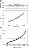
FIGURE 1
FIGURE 1
(a) Sedimentation equilibrium data and analysis at 20°C of a sample made with 80 μM FPK3W peptide in 5 mM pH 7 HEPES buffer. The bottom panel displays the 280-nm absorbance as a function of centrifugal radius. The data are represented as open circles and were obtained after 20 h spinning at 52,000 rpm. The superimposed curve represents the best fit to the data and was obtained using a molar mass of 2600 g, which is comparable to the 2700-g molar mass of a peptide monomer. The upper panel shows the differences between the experimental and best-fit absorbances. (b) Sedimentation equilibrium data at 5000 rpm of a sample made with 80 μM FPW peptide in 5 mM pH 7 HEPES buffer. The logarithm of 280-nm absorbance is plotted as a function of (radius)2. The data are represented as dark circles and show significant curvature, which indicates that the peptide does not form a single oligomeric species. The dark-dotted and light-dashed lines represent the results that would be expected for 230,000- and 2300-g molar mass species and correspond to FPW 100 mers and monomers, respectively. It appears that the solution contains large FPW oligomers.
FIGURE 2
FIGURE 2
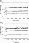
Lipid mixing induced in 100-nm diameter LM3 vesicles by fusion peptides. The total lipid and cholesterol concentrations were 150 and 75 μM, respectively, and the same batches of vesicles were used for all assays. In a and b, the curves correspond to the following peptides: (i) FP; (ii) FPK3; (iii) FPK3W; and (iv) FPW. In a, the peptide concentration was 1.5 μM, and in b the peptide concentration was 3.0 μM. Data acquisition began after manual addition and mixing of the peptide in the vesicle solution. Because the time for these steps is significant relative to characteristic lipid mixing times, the apparent lipid mixing is nonzero at 0 s.
FIGURE 3
FIGURE 3
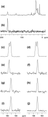
13C solid-state NMR spectra of fusion peptide samples. Spectrum a is the unfiltered S0 spectrum of an FPK3/LM3 sample containing ∼0.4 μmol peptide, 40 μmol total lipid, and 20 μmol cholesterol, and spectrum b is the REDOR-filtered difference spectrum of this sample. Because the peptide was 13C carbonyl labeled at Phe-8 and 15N labeled at Leu-9, the difference spectrum is dominated by the Phe-8 carbonyl signal. The vertical scale in spectrum b is expanded by a factor of ∼6.5 relative to the scale in spectrum a. In cj, REDOR-filtered difference spectra are displayed for samples containing different fusion peptide constructs: (c) FP freeze-dried from a ∼500-μM aqueous solution; (d) FPK3 freeze-dried from a ∼100-μM aqueous solution; (e) FPK3 (2 mM) in dodecylphosphocholine detergent solution (200 mM); (f) FP, (g and j) FPK3, (h) FPW, and (i) FPK3W associated with LM3 (∼0.4 μmol peptide, 40 μmol total lipid, and 20 μmol cholesterol). The samples used for spectra fi were made by method 1, with mixing of aqueous peptide and vesicle solutions. The sample used for spectrum j was made by method 2, with organic cosolubilization of peptide, lipid, and cholesterol, followed by evaporation of the organic solvent and hydration with aqueous buffer. The spectra in c and d display two peaks and indicate conformational heterogeneity for the lyophilized peptide. The spectrum in e is peaked in the 175–176-ppm range, and the spectra in fj are peaked in the 171–172-ppm range. The chemical shifts are consistent with helical structure near Phe-8 for the detergent sample and β-strand structure near Phe-8 for the LM3 samples. Data were acquired using cross-polarization, 8-kHz MAS frequency, and temperatures of −50°C for spectra ad and fj and −80°C for spectrum e. Each spectrum was processed with 50-Hz Gaussian line broadening and polynomial baseline correction. There were 12,288 transients summed in spectrum a, and the total (S0 + S1) numbers of transients in the REDOR-filtered difference spectra were (b) 24,576; (c) 20,000; (d) 31,488; (e) 132,524; (f) 22,336; (g) 24,576; (h) 159,808; (i) 186,560; and (j) 166,336.

Similar articles

Cited by

References

    1. Afonin, S., R. W. Glaser, M. Berditchevskaia, P. Wadhwani, K. H. Guhrs, U. Mollmann, A. Perner, and A. S. Ulrich. 2003. 4-Fluorophenylglycine as a label for 19F NMR structure analysis of membrane-associated peptides. ChemBioChem. 4:1151–1163. - PubMed
    1. Aloia, R. C., H. Tian, and F. C. Jensen. 1993. Lipid composition and fluidity of the human immunodeficiency virus envelope and host cell plasma membranes. Proc. Natl. Acad. Sci. USA. 90:5181–5185. - PMC - PubMed
    1. Balbach, J. J., J. Yang, D. P. Weliky, P. J. Steinbach, V. Tugarinov, J. Anglister, and R. Tycko. 2000. Probing hydrogen bonds in the antibody-bound HIV-1 gp120 V3 loop by solid state NMR REDOR measurements. J. Biomol. NMR. 16:313–327. - PubMed
    1. Bennett, A. E., C. M. Rienstra, M. Auger, K. V. Lakshmi, and R. G. Griffin. 1995. Heteronuclear decoupling in rotating solids. J. Chem. Phys. 103:6951–6958.
    1. Bentz, J. 2000a. Membrane fusion mediated by coiled coils: a hypothesis. Biophys. J. 78:886–900. - PMC - PubMed

Publication types

LinkOut - more resources