Comparative Study
. 2004 Sep 14;101(37):13542-7.
doi: 10.1073/pnas.0405579101. Epub 2004 Sep 2.

Comparative structural modeling and inference of conserved protein classes in Drosophila seminal fluid

Affiliations
Comparative Study

Comparative structural modeling and inference of conserved protein classes in Drosophila seminal fluid

Jacob L Mueller et al. Proc Natl Acad Sci U S A. .

Abstract

The constituents of seminal fluid are a complex mixture of proteins and other molecules, most of whose functions have yet to be determined and many of which are rapidly evolving. As a step in elucidating the roles of these proteins and exposing potential functional similarities hidden by their rapid evolution, we performed comparative structural modeling on 28 of 52 predicted seminal proteins produced in the Drosophila melanogaster male accessory gland. Each model was characterized by defining residues likely to be important for structure and function. Comparisons of known protein structures with predicted accessory gland proteins (Acps) revealed similarities undetectable by primary sequence alignments. The structures predict that Acps fall into several categories: regulators of proteolysis, lipid modifiers, immunity/protection, sperm-binding proteins, and peptide hormones. The comparative structural modeling approach indicates that major functional classes of mammalian and Drosophila seminal fluid proteins are conserved, despite differences in reproductive strategies. This is particularly striking in the face of the rapid protein sequence evolution that characterizes many reproductive proteins, including Drosophila and mammalian seminal proteins.


Figures

Fig. 1.
Fig. 1.
Candidate hormone-binding pocket of CG8137. (A) Superimposed stereoview ribbon diagrams of CG8137 and PCI (cleavage fragment not shown), in the same orientation as the surface potential figures, show the conserved serpin family secondary structures. PCI ribbons are in red and CG8137 ribbons are in yellow, with a green arrow indicating the position of the truncated N-terminal helices forming the hydrophobic pocket. (B) Molecular surface of CG8137 is shown in stereoview with hydrophobic surface areas shown in red and hydrophilic surfaces shown in blue. The yellow arrow indicates the position of the candidate hydrophobic pocket. Glu-42 in CG8137 is primarily responsible for the difference in electrostatic surface potential charge (not shown) between CG8137 and PCI.
Fig. 2.
Fig. 2.
Acid-lipase structure characteristics of CG8093 and active-site comparison of CG8093, CG18284, CG11598, and 1hlg. (A) Complete predicted structure of CG8093 and its predicted CAP-domain in purple, which when removed (Fig. 5, which is published as supporting information on the PNAS web site) exposes the active site (red arrow). (B) Overlay of CG8093 (red), CG18284 (blue), CG11598 (green), and 1hlg (orange) active sites showing similar spatial orientation. The lipase catalytic triad residues Ser, His, and Asp are superimposed to show active site spatial integrity across multiple acid lipase structures. The additional His, adjacent to the Ser, is required for active site stability and is a component of the lipase signature motif, GHSXG. Residue positions are given for CG8093 only.
Fig. 3.
Fig. 3.
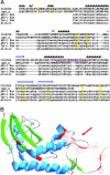
Sequence and structural characteristics of the CRISP domain of CG10284. (A) Sequence alignment of the CRISP domains (entire protein coding regions are not shown) of CG10284 (D. melanogaster), and members of the CRISP family 1qnx_A(Vespula vulgaris), AEG-1 (Mus musculus), AEG-1 (Homo sapiens), TPX-1 (Homo sapiens), and allurin (Xenopus laevis). Residues of known (1qnx_A) and threaded (CG10284) structure are capitalized. CG10284 residue positions 48–283 are shown. Overlapping predicted and known secondary structural regions are depicted above each alignment. α-Helices are colored red, β-strands blue, and 310 helices green. Conserved cysteines are highlighted in yellow. CG10284's region with the excessive amino acid replacements between D. simulans and D. melanogaster is underlined in purple. (B) Overall structure and mapping of amino acid replacements of CG10284 when compared between D. simulans and D. melanogaster. The overall structure of CG10284 threaded onto 1qnx_A (venom antigen) is shown in ribbon representation with α-helices shown in blue and β-strands shown in green. Amino acid replacement sites are shown in red and have been marked by exposing their side chains and electron density clouds. The red arrow points to the loop region where a high frequency of amino acid replacements exists between D. simulans and D. melanogaster.

Similar articles

Cited by

References

    1. Swanson, W. J., Clark, A. G., Waldrip-Dail, H. M., Wolfner, M. F. & Aquadro, C. F. (2001) Proc. Natl. Acad. Sci. USA 98, 7375–7379. - PMC - PubMed
    1. Wolfner, M. F. (2002) Heredity 88, 85–93. - PubMed
    1. Wolfner, M. F., Harada, H. A., Bertram, M. J., Stelick, T. J., Kraus, K. W., Kalb, J. M., Lung, Y. O., Neubaum, D. M., Park, M. & Tram, U. (1997) Insect Biochem. Mol. Biol. 27, 825–834. - PubMed
    1. Monsma, S. A., Harada, H. A. & Wolfner, M. F. (1990) Dev. Biol. 142, 465–475. - PubMed
    1. Chen, P. S., Stumm-Zollinger, E., Aigaki, T., Balmer, J., Bienz, M. & Bohlen, P. (1988) Cell 54, 291–298. - PubMed

Publication types

MeSH terms

LinkOut - more resources