

Application of Doppler technology as an aid in identifying vascular structures during laparoscopy
- PMID: 15347115
- PMCID: PMC3016804
Application of Doppler technology as an aid in identifying vascular structures during laparoscopy
Abstract
Background: Intraoperative ultrasound has been used extensively during open surgery to assess bowel viability, to identify vascular structures, and to assess for congenital abnormalities. The extension of this technology in laparoscopic procedures has been hampered by the size of the equipment and the significant learning curve that accompanies its use.
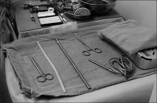
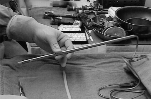
Methods: Using a readily available Parks Inst. Co. Doppler Probe (8.1 MHz) and a 15-inch section of thick-walled, 9.5-mm OD Stainless Steel tubing, a Laparoscopic Doppler Probe was constructed. The parts were separately gas-sterilized, and a small segment of Penrose drain was used to create an airtight seal. The probe was passed through a 10-mm port, allowing assessment of vascular structures.
Results: Two Laparoscopic Doppler Probes were available for evaluation during a 1-month period at our hospital. Surgeons were then surveyed at the end of the 1-month period as to the utility of the devices.
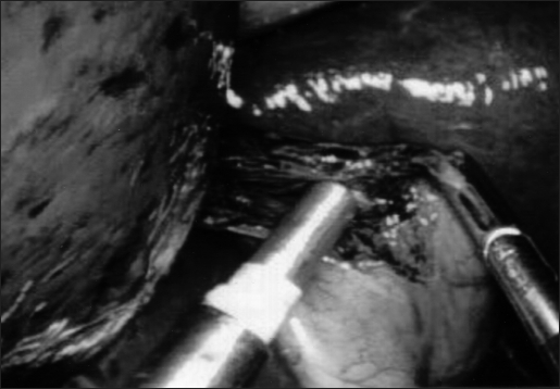
Conclusions: The Laparoscopic Doppler probe was used to identify the cystic artery during gallbladder dissection, to assess mesenteric blood vessels during laparoscopic colectomy, and to identify femoral vessels during laparoscopic preperitoneal hernia repair. It was found to be quick to construct, easy to use, and provided useful information to the operating surgeon.
Figures
References
-
- Hartley JE, Kumar H, Drew PJ, et al. Laparoscopic ultrasound for the detection of hepatic metastases during laparoscopic colorectal cancer surgery. Dis Colon Rectum. 2000;43(3):320–324 - PubMed
-
- Tsioulias GJ, Wood TF, Chung MH, Morton DL, Bilchik A. Diagnostic laparoscopy and laparoscopic ultrasonography optimize the staging and resectability of intraabdominal neoplasms. Surg Endosc. 2001;15(9):1016–1019 - PubMed
-
- Catheline J, Rizk N, Champault G. A comparison of laparoscopic ultrasound versus cholangiography in the evaluation of the biliary tree during laparoscopic cholecystectomy. Eur J Ultrasound. 1999;10(1):1–9 - PubMed
-
- Tomonaga T, Filipi CJ, Lowham A, Martinez T. Laparoscopic intracorporeal ultrasound cystic duct length measurement: a new technique to prevent common bile duct injuries. Surg Endosc. 1999;13(2):183–185 - PubMed
MeSH terms
LinkOut - more resources
Full Text Sources