Comparative Study
. 2004 Nov;87(5):3193-204.
doi: 10.1529/biophysj.104.048447. Epub 2004 Sep 3.

Distinct effects on Ca2+ handling caused by malignant hyperthermia and central core disease mutations in RyR1

Affiliations
Comparative Study

Distinct effects on Ca2+ handling caused by malignant hyperthermia and central core disease mutations in RyR1

Robert T Dirksen et al. Biophys J. 2004 Nov.

Abstract

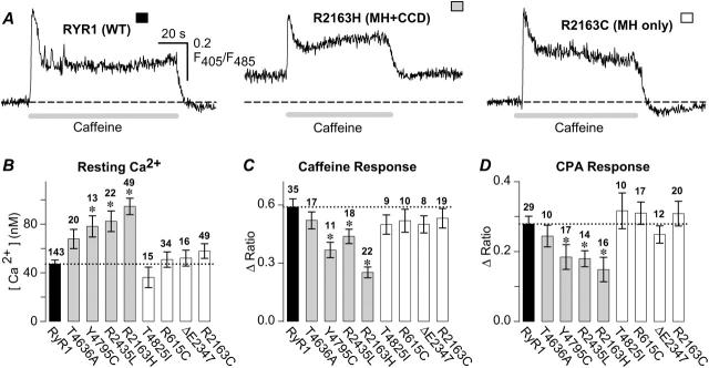
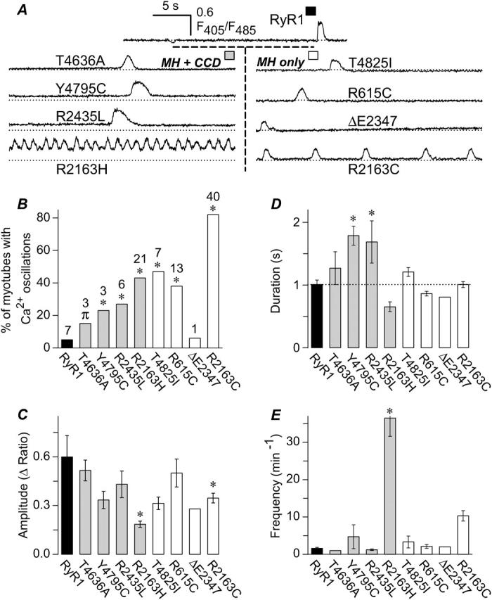
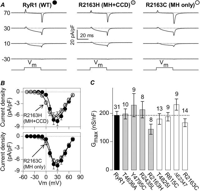
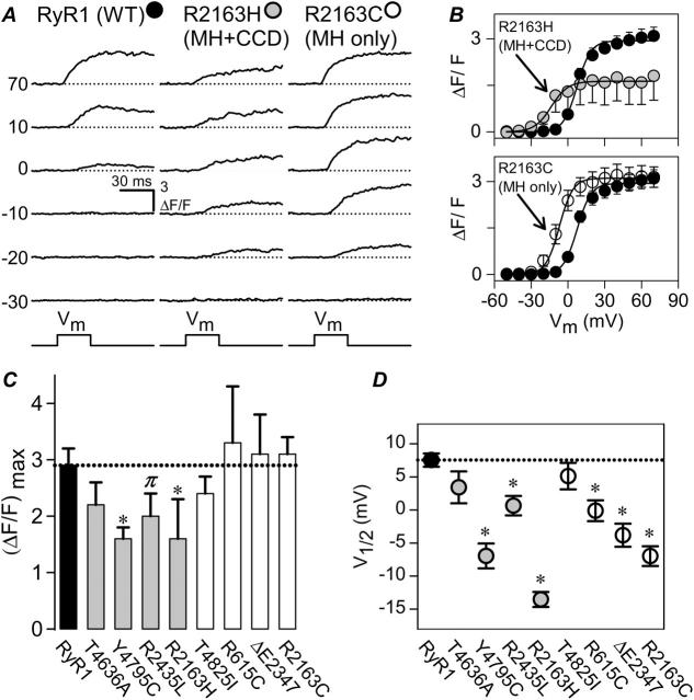
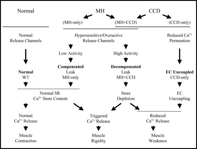
Malignant hyperthermia (MH) and central core disease (CCD) are disorders of skeletal muscle Ca2+ homeostasis that are linked to mutations in the type 1 ryanodine receptor (RyR1). Certain RyR1 mutations result in an MH-selective phenotype (MH-only), whereas others result in a mixed phenotype (MH + CCD). We characterized effects on Ca2+ handling and excitation-contraction (EC) coupling of MH-only and MH + CCD mutations in RyR1 after expression in skeletal myotubes derived from RyR1-null (dyspedic) mice. Compared to wild-type RyR1-expressing myotubes, MH + CCD- and MH-only-expressing myotubes exhibited voltage-gated Ca2+ release (VGCR) that activated at more negative potentials and displayed a significantly higher incidence of spontaneous Ca2+ oscillations. However, maximal VGCR was reduced only for MH + CCD mutants (Y4795C, R2435L, and R2163H) in which spontaneous Ca2+ oscillations occurred with significantly longer duration (Y4795C and R2435L) or higher frequency (R2163H). Notably, myotubes expressing these MH + CCD mutations in RyR1 exhibited both increased [Ca2+]i and reduced sarcoplasmic reticulum (SR) Ca2+ content. We conclude that MH-only mutations modestly increase basal release-channel activity in a manner insufficient to alter net SR Ca2+ content ("compensated leak"), whereas the mixed MH + CCD phenotype arises from mutations that enhance basal activity to a level sufficient to promote SR Ca2+ depletion, elevate [Ca2+]i, and reduce maximal VGCR ("decompensated leak").


Figures

FIGURE 1
FIGURE 1
MH + CCD mutations in RyR1 induce parallel and opposing changes in resting Ca2+ and releasable SR Ca2+ content. (A) Representative caffeine-induced Ca2+ responses obtained from intact Indo-1 AM-loaded dyspedic myotubes expressing WT RyR1, R2163H (MH + CCD), or R2163C (MH-only). Intracellular Ca2+ levels were recorded in the absence (resting Ca2+) and presence of 10 mM caffeine (shaded bar). For each panel, a dashed line representing the average resting fluorescence ratio for WT RyR1-expressing myotubes is shown for comparison. (B) Average resting Ca2+ levels for dyspedic myotubes expressing either WT RyR1 (black bar), four different MH + CCD mutant RyR1s (shaded bars), and four different MH-only mutant RyR1s (white bars). Number of experiments is given above each bar. (C and D) Average peak responses to application of maximal concentrations of caffeine (C, 10 mM) and CPA (D, 30 μM) in dyspedic myotubes expressing wild-type RyR1 (black bar) and each of the MH + CCD (shaded bars) and MH-only (white bars) mutations in RyR1 illustrated in B. A one-way ANOVA analysis (p < 0.05) revealed that mean WT RyR1 data in BD were statistically indistinguishable from the corresponding MH-only group data, whereas MH + CCD group data were statistically different from both WT RyR1 and MH-only group data. Asterisks indicate significant differences compared to WT RyR1 using ANOVA/Dunnett analysis (P < 0.05). In this and subsequent figures, RyR1 mutants for each group (MH + CCD and MH-only) are presented in increasing order for elevating resting Ca2+ as shown in B.
FIGURE 2
FIGURE 2
MH-only- and MH + CCD-expressing dyspedic myotubes exhibit increased incidence of spontaneous fluctuations in intracellular Ca2+ (Ca2+ oscillations). (A) Representative spontaneous Ca2+ oscillations in intact Indo-1 AM-loaded myotubes expressing WT RyR1 (top) and each of the eight different MH + CCD (left) and MH-only mutants (right). For comparison, a dashed line representing the average resting fluorescence ratio for WT RyR1-expressing myotubes is shown for each construct. (B) Global intracellular Ca2+ levels were continuously recorded for all of the cells analyzed in Fig. 1 B and spontaneous Ca2+ oscillations were analyzed for the percentage of myotubes exhibiting Ca2+ oscillations during the first 60 s of recording (numbers above each bar represent the number of myotubes from Fig. 1 B that exhibited spontaneous oscillations for each construct). Statistical significance in the percentage MH + CCD- and MH-only-expressing myotubes exhibiting spontaneous Ca2+ oscillations compared to that of WT RyR1-expressing myotubes was assessed using a nonparametric chi-squared test (*p < 0.05, πp < 0.1). (CE) Spontaneous Ca2+ oscillation amplitude (C), duration (D), and frequency (E) analyzed from WT RyR1-, MH + CCD-, and MH-only-expressing myotubes. Asterisks indicate significant differences compared to RyR1 using ANOVA/Dunnett analysis (P < 0.05). MH/CCD mutations significantly increased the duration (Y4795C and R2435L) and frequency (R2163H) of spontaneous Ca2+ oscillations, whereas both MH-only and MH/CCD mutations in R2163 (R2163C and R2163H) resulted in a significant reduction in Ca2+ oscillation amplitude.
FIGURE 3
FIGURE 3
The MH + CCD and MH-only mutations in RyR1 fully restore retrograde coupling with sarcolemmal DHPRs. (A) Representative whole-cell L-type Ca2+ currents recorded from RyR1- (left), R2163H- (middle), and R2163C-expressing (right) dyspedic myotubes. Current traces were elicited in response to brief (30-ms) membrane depolarizations to the indicated membrane potentials. Dashed lines represent the zero current level. (B) Average (±SEM) peak current-voltage (I-V) relationships for dyspedic myotubes expressing wild-type RyR1 (black circles; n = 31), R2163H (shaded circles, top; n = 8), and R2163C (open circles, bottom; n = 14). The average values (±SEM) for the parameters obtained by fitting each myotube within a group separately to Eq. 1 are given in Table 1 (I-V data). The solid lines through the data were obtained using Eq. 1 and the corresponding parameters given in Table 1 (I-V data). (C) Comparison of maximal L-channel conductance (Gmax) obtained from RyR1- (black bar), MH + CCD- (shaded bars), and MH-only-expressing (white bars) myotubes. Number of experiments is given above each bar.
FIGURE 4
FIGURE 4
Voltage-gated SR Ca2+ release is preferentially reduced in MH + CCD-expressing myotubes. (A) Representative intracellular Ca2+ transients elicited by 30-ms test pulses to the indicated potentials. Representative Ca2+ transients are displayed for dyspedic myotubes expressing RyR1 (left), R2163H (middle), and R2163C (right). Dashed lines represent basal fluorescence before depolarization. (B) Average voltage-dependence of intracellular Ca2+ transients (measured at the end of each test pulse) for dyspedic myotubes expressing wild-type RyR1 (black circles; n = 31), R2163H (shaded circles, top; n = 8), and R2163C (open circles, bottom; n = 14). The average values (±SEM) for the parameters obtained by fitting each myotube within a group separately to Eq. 2 are given in Table 1 (ΔF/F-V data). The solid lines through the data were obtained using Eq. 2 and the corresponding parameters given in Table 1 (ΔF/F-V data). For clarity, average values (±SEM) for maximal voltage-gated SR Ca2+ release ([ΔF/F]max) and the voltage required for half-maximal activation of release (VF1/2) for WT RyR1 (black bar/symbol), MH + CCD mutant RyR1s (shaded bars/symbols), and MH-only mutant RyR1s (white bars/symbols) are plotted in C and D, respectively. Dashed lines in C and D correspond to mean data for WT RyR1. *p < 0.05, πp < 0.1 compared to RyR1.
FIGURE 5
FIGURE 5
Schematic depicting fundamentally distinct cellular consequences of mutations in RyR1 that result in MH-only (compensated leak), MH + CCD (decompensated leak), and CCD-only (EC uncoupled) mutations in RyR1. In this scheme, MH in the absence of CCD arises from RyR1 mutations that result in overactive/supersensitive release channels that fail to cause a net change in steady-state SR Ca2+ content (compensated leak), but predispose muscle to increased MHS upon exposure to triggering agents. The primary distinguishing feature of CCD mutations is a reduction in maximal voltage-gated release that results either from a net reduction in steady-state SR Ca2+ content or EC uncoupling (functional uncoupling of depolarization from the release of Ca2+ from a full SR store). MH and CCD coincidence occurs for mutations in RyR1 that enhance RyR1 activity/sensitivity to an extent sufficient to result in a partial depletion of SR Ca2+ stores decompensated leak).

References

    1. Avila, G., and R. T. Dirksen. 2000. Functional impact of the ryanodine receptor on the skeletal muscle L-type Ca2+ channel. J. Gen. Physiol. 115:467–480. - PMC - PubMed
    1. Avila, G., and R. T. Dirksen. 2001. Functional effects of central core disease mutations in the cytoplasmic region of the skeletal muscle ryanodine receptor. J. Gen. Physiol. 118:277–290. - PMC - PubMed
    1. Avila, G., E. H. Lee, C. F. Perez, P. D. Allen, and R. T. Dirksen. 2003a. FKBP12 binding to RyR1 modulates excitation-contraction coupling in mouse skeletal myotubes. J. Biol. Chem. 278:22600–22608. - PubMed
    1. Avila, G., J. J. O'Brien, and R. T. Dirksen. 2001. Excitation-contraction uncoupling by a human central core disease mutation in the ryanodine receptor. Proc. Natl. Acad. Sci. USA. 98:4215–4220. - PMC - PubMed
    1. Avila, G., K. M. S. O'Connell, and R. T. Dirksen. 2003b. The pore region of the skeletal muscle ryanodine receptor is a primary locus for excitation-contraction uncoupling in central core disease. J. Gen. Physiol. 121:277–286. - PMC - PubMed

Publication types

MeSH terms

Substances