Comparative Study
. 2004 Nov;87(5):3336-43.
doi: 10.1529/biophysj.104.040634. Epub 2004 Sep 3.

Cholesterol depletion increases membrane stiffness of aortic endothelial cells

Affiliations
Comparative Study

Cholesterol depletion increases membrane stiffness of aortic endothelial cells

Fitzroy J Byfield et al. Biophys J. 2004 Nov.

Abstract

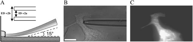
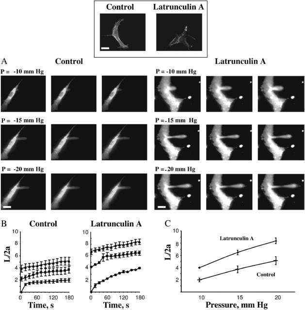
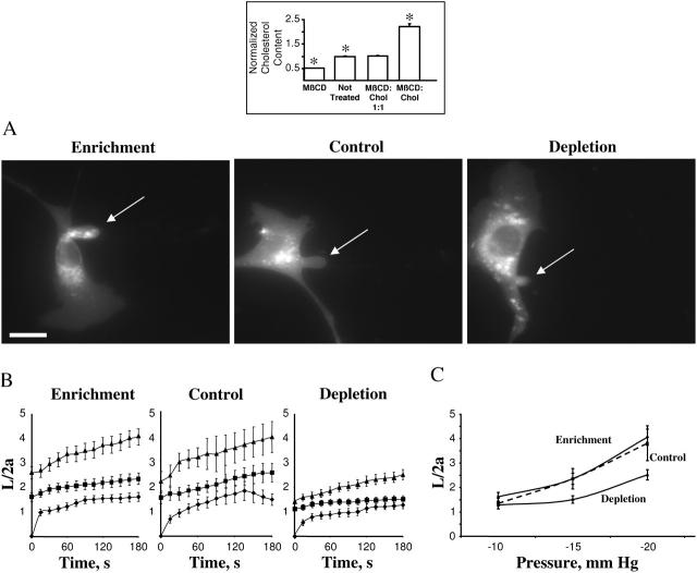
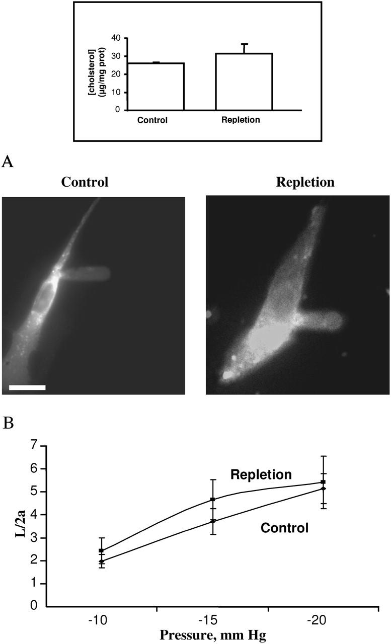
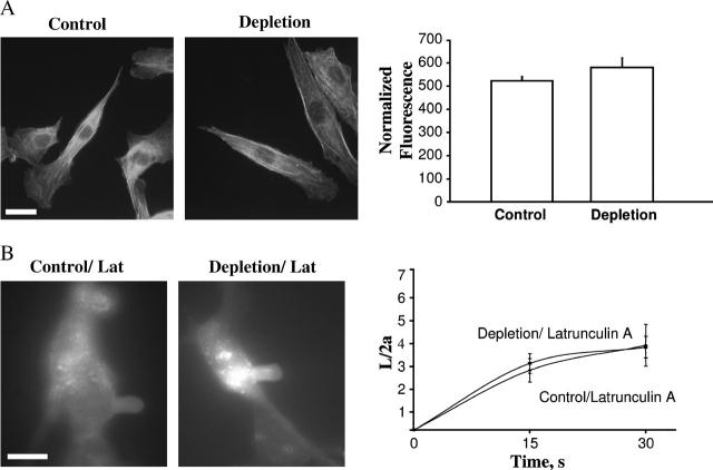
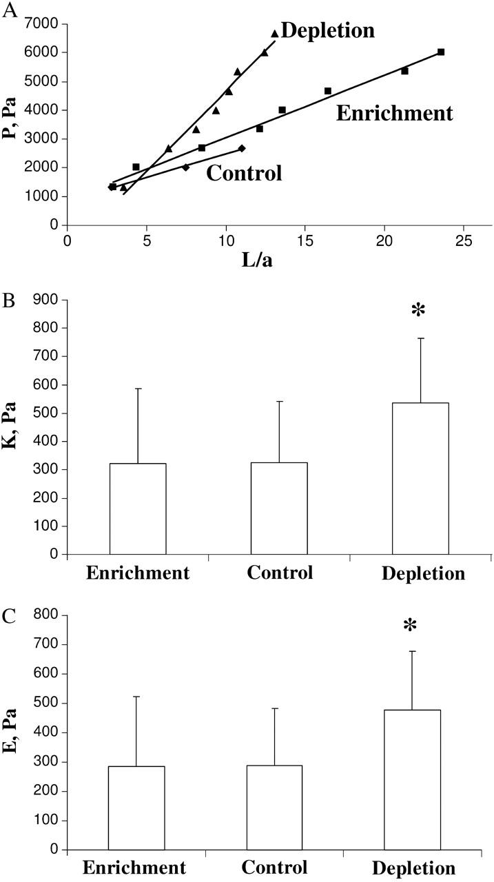
This study has investigated the effect of cellular cholesterol on membrane deformability of bovine aortic endothelial cells. Cellular cholesterol content was depleted by exposing the cells to methyl-beta-cyclodextrin or enriched by exposing the cells to methyl-beta-cyclodextrin saturated with cholesterol. Control cells were treated with methyl-beta-cyclodextrin-cholesterol at a molar ratio that had no effect on the level of cellular cholesterol. Mechanical properties of the cells with different cholesterol contents were compared by measuring the degree of membrane deformation in response to a step in negative pressure applied to the membrane by a micropipette. The experiments were performed on substrate-attached cells that maintained normal morphology. The data were analyzed using a standard linear elastic half-space model to calculate Young elastic modulus. Our observations show that, in contrast to the known effect of cholesterol on membrane stiffness of lipid bilayers, cholesterol depletion of bovine aortic endothelial cells resulted in a significant decrease in membrane deformability and a corresponding increase in the value of the elastic coefficient of the membrane, indicating that cholesterol-depleted cells are stiffer than control cells. Repleting the cells with cholesterol reversed the effect. An increase in cellular cholesterol to a level higher than that of normal cells, however, had no effect on the elastic properties of bovine aortic endothelial cells. We also show that although cholesterol depletion had no apparent effect on the intensity of F-actin-specific fluorescence, disrupting F-actin with latrunculin A abrogated the stiffening effect. We suggest that cholesterol depletion increases the stiffness of the membrane by altering the properties of the submembrane F-actin and/or its attachment to the membrane.


Figures

FIGURE 1
FIGURE 1
Micropipette aspiration technique for substrate attached BAECs. (A) Schematic side view of a micropipette approaching a substrate-attached cell. Inset, depiction of micropipette parameters used in analysis, where ID = 2a = internal diameter and ED = 2b = external diameter. (B) Bright contrast image of a micropipette shank touching a typically shaped cell used in aspiration experiments. (C) Fluorescent image of the same cell labeled with DiIC18. The micropipette is still present but is invisible. Scale bar, 30 μm.
FIGURE 2
FIGURE 2
Membrane deformation of substrate-anchored endothelial cells under control conditions and after being exposed to latrunculin A. (Inset) The effect of latrunculin A on F-actin filaments visualized with rhodamine phalloidin. The bright spots in phalloidin staining were observed in most of the images suggesting that the treatment may result in collapse and condensation of actin fibers. The bar is 25 μm. (A) Progressive deformation of control cells compared to that of cells exposed to 2μM latrunculin A for 10 minutes. The edges of the cells have dimmer fluorescence than cell centers because in the cell center fluorescent signal comes not only from the plasma membrane but also from endocytosed vesicles. (B) Time course of membrane deformation where L is the aspirated length of the membrane projection and 2a is the inner diameter of the pipette. Cells were aspirated at −10 mm Hg (♦), −15 mm Hg (▪), and −20 mm Hg (▴). (C) Maximal membrane deformation as a function of applied pressure. Maximal deformation was determined by taking the values at which the aspiration length plateaued for each pressure. The maximal normalized length in latrunculin treated cells was significantly greater than control cells for all pressures (P < 0.05).
FIGURE 3
FIGURE 3
Effect of cellular cholesterol levels on membrane deformation of BAECs. (Inset) Effects of MβCD and MβCD-cholesterol on the levels of free cholesterol in BAECs. (A) Typical images of membrane deformation of cholesterol-enriched, cholesterol-depleted, and control cells (control cells were exposed to MβCD/MβCD-cholesterol mixture at 1:1 ratio that had no effect on the level of free cholesterol in the cells (see inset). The images shown depict the maximal deformation at −15 mm Hg. The arrow indicates the position of the aspirated projection. The bar is 30 μm. (B) Average time courses of aspirated lengths for the three experimental cell populations. (C) Maximal aspirated lengths plotted as a function of the applied pressure. The maximal normalized length in depleted cells was significantly lower than that of control cells for pressures −15 mm Hg and −20 mm Hg (P < 0.05).
FIGURE 4
FIGURE 4
Effect of repletion on membrane deformation. (Inset) Free cholesterol level of cells after cholesterol repletion compared to that of control cells. (A) Typical images of the maximal deformation of a typical repleted cell (left) compared to that of a control cell (right) at −15 mm Hg. The bar is 25 μm. (B) Maximal normalized aspirated length as a function of pressure for repleted and control cells. Repleted cells (n = 12), Control cells (n = 14). There was statistically no difference between the two conditions.
FIGURE 5
FIGURE 5
The role of F-actin in the stiffening effect of cholesterol depletion. (A) Effect of cholesterol depletion on F-actin specific staining. (Left panel) Rhodamine-phalloidin labeling of F-actin in control cells and cholesterol-depleted cells. (Right panel) Average fluorescence intensity of rhodamine phalloidin-labeled F-actin in control (n = 20) and depleted cells (n = 20) in a single experiment. There was no significant difference in intensity between depleted and control cells. Identical results were obtained in four independent experiments. (B) Effect of latrunculin A on the membrane deformation of control and cholesterol-depleted cells. (Left panel) Images of maximal deformation of a control and a cholesterol-depleted cell after being treated with latrunculin A. These images were acquired after 30 s at −5 mm Hg. The bar is 30 μm. (Right panel) Average time courses of control (n = 5) and depleted cells (n = 4) aspirated at −5 mm Hg. Only 30 s of the time course was analyzed because most cells broke beyond this point.
FIGURE 6
FIGURE 6
Analysis of the elastic properties of BAECs under different cholesterol conditions. (A) Typical data of the dimensionless aspirated projection length, L/a, as a function of the negative applied pressure difference, P, for control, cholesterol-enriched, and cholesterol-depleted cells with their respective least-squares fit (solid lines). (B) Elastic modulus K for control (7 cells), enrichment (10 cells), and depletion (17 cells) as obtained by least-squares fit of a straight line to the data. Depleted cells are significantly stiffer (P < 0.05) than control and cholesterol-enriched cells. Mean ± SE. (C) Young's modulus E according to the homogeneous half-space model (Eq. 1) for the data shown in B. Depleted cells have a significantly larger Young's modulus (P < 0.05) than control and cholesterol-enriched cells. Mean ± SE.

References

    1. Brener, E., S. Rubinstein, G. Cohen, K. Shternall, J. Rivlin, and H. Breitbart. 2003. Remodeling of the actin cytoskeleton during mammalian sperm capacitation and acrosome reaction. Biol. Reprod. 68:837–845. - PubMed
    1. Brown, A. D., and E. London. 2000. Structure and function of sphingolipid- and cholesterol-rich membrane rafts. J. Biol. Chem. 275:17221–17224. - PubMed
    1. Brulet, P., and H. M. McConnell. 1976. Lateral hapten mobility and immunochemistry of model membranes. Proc. Natl. Acad. Sci. USA. 73:2977–2981. - PMC - PubMed
    1. Caroni, P. 2001. New EMBO members' review: actin cytoskeleton regulation through modulation of PI(4,5)P(2) rafts. EMBO J. 20:4332–4336. - PMC - PubMed
    1. Chien, S., K. L. Sung, R. Skalak, S. Usami, and A. Tozeren. 1978. Theoretical and experimental studies on viscoelastic properties of erythrocyte membrane. Biophys. J. 24:463–487. - PMC - PubMed

Publication types