. 2004 Sep 14;101(37):13648-53.
doi: 10.1073/pnas.0405310101. Epub 2004 Sep 3.

Behavioral and regulatory abnormalities in mice deficient in the NPAS1 and NPAS3 transcription factors

Affiliations

Behavioral and regulatory abnormalities in mice deficient in the NPAS1 and NPAS3 transcription factors

Claudia Erbel-Sieler et al. Proc Natl Acad Sci U S A. .

Abstract

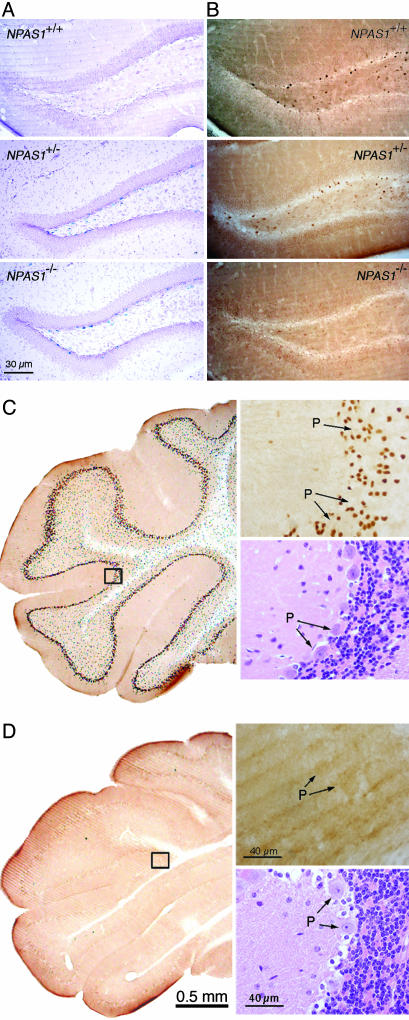
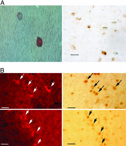
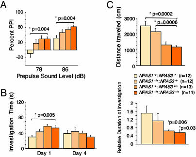
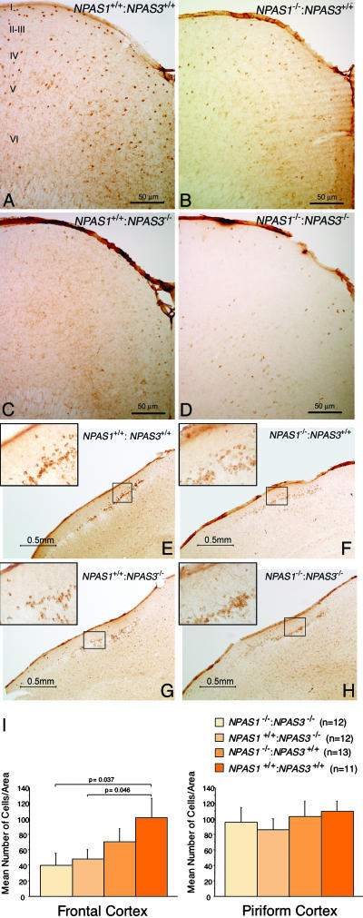
Laboratory mice bearing inactivating mutations in the genes encoding the NPAS1 and NPAS3 transcription factors have been shown to exhibit a spectrum of behavioral and neurochemical abnormalities. Behavioral abnormalities included diminished startle response, as measured by prepulse inhibition, and impaired social recognition. NPAS1/NPAS3-deficient mice also exhibited stereotypic darting behavior at weaning and increased locomotor activity. Immunohistochemical staining assays showed that the NPAS1 and NPAS3 proteins are expressed in inhibitory interneurons and that the viability and anatomical distribution of these neurons are unaffected by the absence of either transcription factor. Adult brain tissues from NPAS3- and NPAS1/NPAS3-deficient mice exhibited a distinct reduction in reelin, a large, secreted protein whose expression has been reported to be attenuated in the postmortem brain tissue of patients with schizophrenia. These observations raise the possibility that a regulatory program controlled in inhibitory interneurons by the NPAS1 and NPAS3 transcription factors may be either substantively or tangentially relevant to psychosis.


Figures

Fig. 1.
Fig. 1.
Samples of NPAS1 and NPAS3 expression in the adult mouse brain. (A) Patterns of β-galactosidase enzyme activity in coronal sections of brain tissue derived from wt (Top), NPAS1+/– (Middle), and NPAS1–/– (Bottom) animals. (B) Staining patterns of matching tissue samples with antibodies prepared against recombinant NPAS1 protein. NPAS1-positive cells are observed in wt (Top) and NPAS1+/– (Middle) samples, but absent in the NPAS1–/– sample (Bottom). (C) Coronal sections of the cerebellum from a wt mouse immunohistochemically processed with the antibody to NPAS3. NPAS3-positive neurons are seen in the granular layer of the cerebellum immediately outside of the Purkinje (P) cell layer. (Upper Right) Shown is the high-magnification image of the boxed area (Left), revealing nuclear, antibody-reactive material. (Lower Right) Shown is hematoxylin/eosin staining of an analogous region of the cerebellum prepared from a wt mouse. (D) Coronal section as in C but from an NPAS3–/– mouse. Despite the absence of antibody-reactive material in the cerebellum of the NPAS3–/– sample (Upper Right), the hematoxylin/eosin staining pattern (Lower Right) could not be distinguished from wt.
Fig. 2.
Fig. 2.
NPAS1 and NPAS3 neurons colocalize with GABA. (A) Photomicrographs of brain sections subjected to double-labeling immunocytochemistry for NPAS1 and GABA (Left) and NPAS1 and GAD-67 (Right). (Left) The 10-μm paraffin section was taken from the hippocampus of a wt mouse. The NPAS1 reaction product in the nucleus was detected with the Vector DAB peroxidase substrate, whereas the blue-gray GABA reaction product in the cytoplasm was detected with the Vector SG peroxidase substrate. (Magnification, ×700.) (Right) This micrograph is a 40-μm vibratome section through the cerebral cortex of an NPAS1–/– mouse stained for β-galactosidase. The blue, nuclear reaction product marks the presence of NPAS1. The section was then exposed to anti-GAD-67 (Chemicon, 1:1,500) followed by the Vector DAB peroxidase substrate to yield a brown, cytoplasmic reaction product. (Calibration bar, 20 μm.) (B) Photomicrographs from the CA1 (Upper) and CA3 (Lower) areas of the hippocampus from a wt mouse taken under fluorescent and bright-field illumination. The nuclear NPAS3 brown reaction product was detected with the Vector DAB peroxidase substrate kit. The fluorescent GAD-67-expressing neurons were detected by using Texas Red Avidin D (Vector Laboratories). Arrows indicate double-labeled neurons. (Calibration bar, 50 μm.)
Fig. 3.
Fig. 3.
Aberrant behavioral patterns in mice deficient in NPAS1 and NPAS3. (A) Impaired prepulse inhibition (PPI) in NPAS1–/–:NPAS3–/– adult mice. Animals were assayed for prepulse inhibition at two prepulse sound levels, 78 and 86 dB. Statistically significant impairment for prepulse inhibition was observed at both sound levels only for double-null animals. (B) Impaired social recognition in NPAS3–/– and NPAS1–/–:NPAS3–/– adult mice. Animals were assayed for social recognition by comparing the investigation time of unfamiliar juveniles on day 1 and reinvestigation on day 4. (Left) Double-null animals showed a statistically significant reduction in initial investigation time. (Right) Both NPAS3 homozygotes and double-null animals failed to display statistically significant social recognition when comparing investigation times on days 1 and 4. (C) Enhanced open-field locomotor activity in NPAS3–/– and NPAS1–/–:NPAS3–/– adult mice. Animals were assayed for open-field locomotor activity. NPAS3 homozygotes and double-null animals showed a statistically significant increase in total distance traveled compared with NPAS1 homozygotes and wt littermates. Thigmotactic analysis revealed that extra locomotor activity took place in the outer rim of the open-field cage.
Fig. 4.
Fig. 4.
Attenuation in reelin expression in brain tissue derived from NPAS1+/+: NPAS3–/– and NPAS1–/–:NPAS3–/– adult mice. Coronal sections of adult mouse brain tissue were prepared from wt (A and E), NPAS1 homozygotes (B and F), NPAS3 homozygotes (C and G), and double-null (D and H) animals. Sections were stained with antibodies specific to reelin. Shown are antibody staining patterns on matching coronal sections of the frontal (AD) and piriform (EH) cortex. Irrespective of brain region, reelin-positive antibody staining was uniformly restricted to the cytoplasm of inhibitory interneurons. Significant attenuation in reelin antibody staining was observed in the frontal cortex of NPAS3 homozygotes and double-null animals. The roman numerals in A designate cortical layers I–VI. (I) Quantitative assessment of reelin expression in adult brain tissue derived from wt, NPAS1+/+:NPAS31–/–, NPAS1–/–: NPAS3+/+, and NPAS1–/–:NPAS3–/– mice. Coronal sections were prepared from matching regions of cerebral and piriform cortex of at least five animals of the four genotypes under study. Sections were stained with antibodies specific to reelin, photographed, and quantitated over equivalent 1.6-mm2 regions for reelin-positive neurons. Observers were blinded to genotype.

References

    1. Zhou, Y.-D., Barnard, M., Tian, H., Li, X., Ring, H.Z., Francke, U., Shelton, J., Richardson, J., Russell, D. W. & McKnight, S. L. (1997) Proc. Natl. Acad. Sci. USA 94, 713–718. - PMC - PubMed
    1. Brunskill, E. W., Witte, D. P., Shreiner, A. B. & Potter, S. S. (1999) Mech. Dev. 88, 237–241. - PubMed
    1. Dudley, C. A., Erbel-Sieler, C., Estill, S. J., Reick, M., Franken, P., Pitts, S. & McKnight, S. L. (2003) Science 301, 379–383. - PubMed
    1. Rutter, J., Reick, M., Wu, L. C. & McKnight, S. L. (2001) Science 293, 510–514. - PubMed
    1. Taylor, B. L. & Zhulin, I. B. (1999) Microbiol. Mol. Biol. Rev. 63, 479–506. - PMC - PubMed

Publication types

MeSH terms