. 2004 Sep 21;101(38):13933-8.
doi: 10.1073/pnas.0404768101. Epub 2004 Sep 3.

Identification of key phosphorylation sites in the circadian clock protein KaiC by crystallographic and mutagenetic analyses

Affiliations

Identification of key phosphorylation sites in the circadian clock protein KaiC by crystallographic and mutagenetic analyses

Yao Xu et al. Proc Natl Acad Sci U S A. .

Abstract

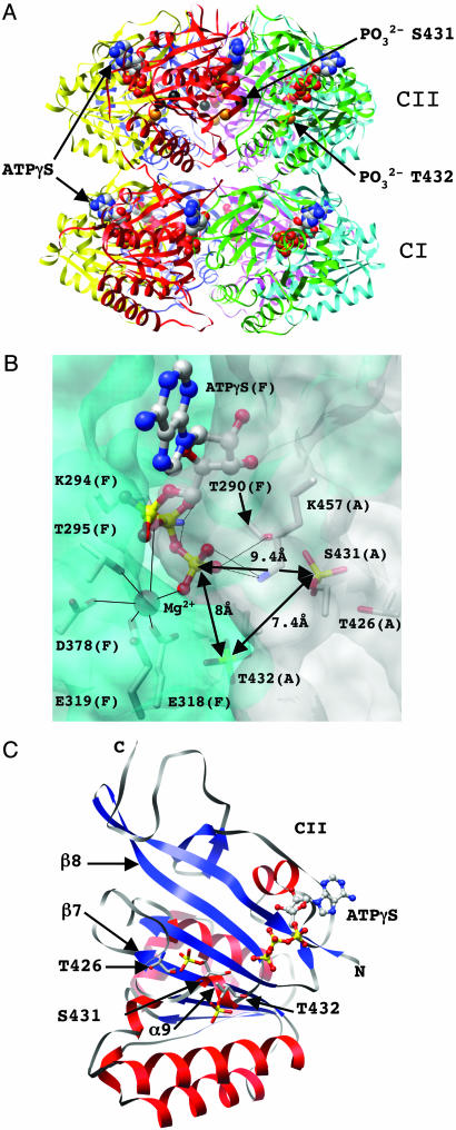
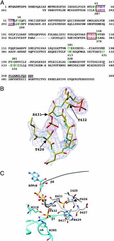
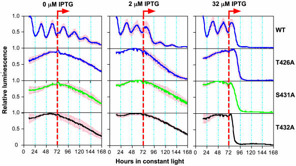
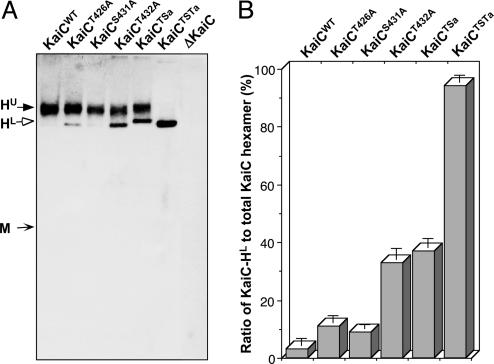
In cyanobacteria, KaiC is an essential hexameric clock protein that forms the core of a circadian protein complex. KaiC can be phosphorylated, and the ratio of phospho-KaiC to non-phospho-KaiC is correlated with circadian period. Structural analyses of KaiC crystals identify three potential phosphorylation sites within a 10-A radius of the ATP binding regions that are at the T432, S431, and T426 residues in the KaiCII domains. When these residues are mutated by alanine substitution singly or in combination, KaiC phosphorylation is altered, and circadian rhythmicity is abolished. These alanine substitutions do not prevent KaiC from hexamerizing. Intriguingly, the ability of KaiC overexpression to repress its own promoter is also not prevented by alanine substitutions at these sites, implying that the capability of KaiC to repress its promoter is not sufficient to allow the clockwork to oscillate. The KaiC structure and the mutational analysis suggest that S431 and T426 may share a phosphate that can shuttle between these two residues. Because the phosphorylation status of KaiC oscillates over the daily cycle, and KaiC phosphorylation is essential for clock function as shown here, daily modulations of KaiC activity by phosphorylation at T432 and S431/T426 seem to be key components of the circadian clockwork in cyanobacteria.


Figures

Fig. 1.
Fig. 1.
Structure of KaiC and phosphorylation sites. (A) Ribbon diagram of the overall structure of the KaiC homo-hexamer with bound ATP molecules shown in a van der Waals representation. The CI and CII domains are labeled, and carbon, oxygen, nitrogen, and phosphorus atoms of ATPs are colored gray, red, blue, and yellow, respectively. Phosphate groups of residues T432 in all subunits are highlighted as orange spheres, and phosphates of residues S431 in subunits A, B, E, and F are highlighted as black spheres. (B) The intersubunit ATP binding site. Portions of the KaiCII domains from adjacent subunits A and F (in parentheses) are shown in a transparent surface representation (colored gray and cyan, respectively). Side chains of T426, S431, and T432, as well as residues interacting (directly or by means of Mg2+) with the triphosphate moiety of ATP, are shown in a stick representation and are labeled. The Mg2+ ion is depicted as a red sphere, and its coordination sphere is indicated with thin solid lines. Distances between the phosphorus atoms of ATP (formula image) and the phosphates of T432 and S431 are highlighted. (C) Secondary and tertiary structure of the KaiCII monomer with the bound ATP molecule. Residues T426, S431, and T432 are located in an extended loop region (gray ribbon) that links the β7 and β8 strands and features a single α-helical turn.
Fig. 2.
Fig. 2.
Consequences of phosphorylation for KaiCII intersubunit organization. (A) Sequence of KaiC from S. elongatus with the KaiCI (top line) and KaiCII (bottom line) domains aligned. Residues 1–13 and 498–519 including the C-terminal His-6 tag (residues 520–525) are disordered in the crystal structure. Amino acids that form part of the Walker A and B motifs are red/blue and boxed; serines and threonines that lie within a 10-Å sphere from the ATP γ-phosphates in the KaiCI and KaiCII domains (P··· Cα distance) are green and are numbered. Serines and threonines contained in the Walker A and B motifs and located within this sphere are blue, and amino acids forming the linker between the CI and CII domains are underlined. (B) Example of the final Fourier 2FoFc sum electron density (blue; 1.5σ level) with the super-imposed FoFc difference electron density (red; 3.5σ level before incorporation of phosphate groups) for S431 and T432 in the F subunit. Only residues 425–433 of the loop region connecting the KaiCII β7 and β8 strands are shown, and selected residues are labeled. (C) Intersubunit interactions as a result of phosphorylation of T432 and S431. Carbon atoms of residues from the A and F subunits are colored gray and cyan, respectively, selected residues and secondary structural elements are labeled, and hydrogen bonds are drawn as thin dashed lines.
Fig. 3.
Fig. 3.
Mutation of key phosphorylation sites alters the phosphorylation status of KaiC. (A) In vivo KaiC phosphorylation profiles in the kaiC-null cyanobacteria (ΔKaiC) expressing trcp-driven wild-type or mutagenized KaiCs (Table 1). After a 3-h pulse of the inducer IPTG in light starting at circadian time 0 (CT0), the expression patterns of KaiC proteins were determined by immunoblotting with mouse anti-KaiC antibody. Filled arrows indicate phosphorylated KaiC bands, and the open arrow denotes nonphosphorylated KaiC bands. (B) Densitometry of the phospho-KaiC levels in various strains, normalized to the density of the phospho-KaiCS431A band. The values are mean ± SD; asterisks denote significant differences vs. wild type (*, P < 0.05; **, P < 0.01).
Fig. 4.
Fig. 4.
Mutation of potential phosphorylation residues of KaiC abolishes circadian rhythmicity. After a 12-h synchronizing dark pulse, colonies of kaiC-null cyanobacteria harboring the constructs for trcp-driven wild-type KaiC (WT) or mutagenized KaiCs (T426A, S431A, or T432A) were released to constant light (LL) for measurement of kaiBCp-driven luminescence. After 72 h in LL, the inducer IPTG (0, 2, or 32 μM) was added as shown by the orange dashed lines. Heavy solid lines indicate the average luminescence traces, and red bars denote the SD (n = 3 for each trace).
Fig. 5.
Fig. 5.
Mutations of key phosphorylation sites do not affect the hexamerization of KaiC but alter the profiles of the KaiC hexamers. (A) Native-gel immunoblotting assay for wild-type or mutagenized KaiCs in cyanobacterial extracts collected at circadian time 3 (CT3). No KaiC monomer was detected in any strain under the conditions described in Materials and Methods. The predicted approximate positions of the KaiC monomer (M) and two distinct hexamer bands (upper = HU, lower = HL) are indicated by arrows. (B) Ratio of the lower hexamer band (HL) to the total hexamer population (HU + HL) as determined by densitometry. Data are mean ± SD.

Comment in

Similar articles

Cited by

References

    1. Ditty, J. L., Williams, S. B. & Golden, S. S. (2003) Annu. Rev. Genet. 37, 513–543. - PubMed
    1. Johnson, C. H. (2004) Curr. Issues Mol. Biol. 6, 103–110. - PubMed
    1. Johnson, C. H. & Egli, M. (2004) Nat. Struct. Mol. Biol. 11, 584–585. - PubMed
    1. Ishiura, M., Kutsuna, S., Aoki, S., Iwasaki, H., Andersson, C. R., Tanabe, A., Golden, S. S., Johnson C. H. & Kondo T. (1998) Science 281, 1519–1523. - PubMed
    1. Williams, S. B., Vakonakis, I., Golden, S. S. & LiWang, A. C. (2002) Proc. Natl. Acad. Sci. USA 99, 15357–15362. - PMC - PubMed

Publication types

Substances

Associated data

LinkOut - more resources