. 2004 Sep 21;101(38):13927-32.
doi: 10.1073/pnas.0403906101. Epub 2004 Sep 3.

Role of KaiC phosphorylation in the circadian clock system of Synechococcus elongatus PCC 7942

Affiliations

Role of KaiC phosphorylation in the circadian clock system of Synechococcus elongatus PCC 7942

Taeko Nishiwaki et al. Proc Natl Acad Sci U S A. .

Abstract

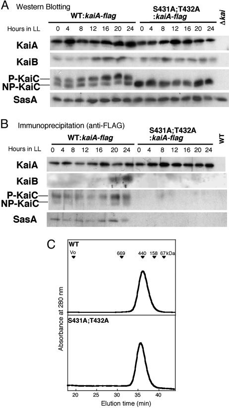
In the cyanobacterium Synechococcus elongatus PCC 7942, KaiA, KaiB, and KaiC are essential proteins for the generation of a circadian rhythm. KaiC is proposed as a negative regulator of the circadian expression of all genes in the genome, and its phosphorylation is regulated positively by KaiA and negatively by KaiB and shows a circadian rhythm in vivo. To study the functions of KaiC phosphorylation in the circadian clock system, we identified two autophosphorylation sites, Ser-431 and Thr-432, by using mass spectrometry (MS). We generated Synechococcus mutants in which these residues were substituted for alanine by using site-directed mutagenesis. Phosphorylation of KaiC was reduced in the single mutants and was completely abolished in the double mutant, indicating that KaiC is also phosphorylated at these sites in vivo. These mutants lost circadian rhythm, indicating that phosphorylation at each of the two sites is essential for the control of the circadian oscillation. Although the nonphosphorylatable mutant KaiC was able to form a hexamer in vitro, it failed to form a clock protein complex with KaiA, KaiB, and SasA in the Synechococcus cells. When nonphosphorylatable KaiC was overexpressed, the kaiBC promoter activity was only transiently repressed. These results suggest that KaiC phosphorylation regulates its transcriptional repression activity by controlling its binding affinity for other clock proteins.


Figures

Fig. 3.
Fig. 3.
The S431A and T432A mutations impair KaiC phosphorylation in vivo and circadian rhythm in Synechococcus.(A) Proteins were extracted from WT and mutant strains at LL4 and LL16 and subjected to immunoblot analysis to examine the phosphorylation state of KaiC. The upper four bands of KaiC from the WT strain are phosphorylated, and the lower two are nonphosphorylated. Only two of the phosphorylated bands were detected from mutant strains containing either S431A or T432A mutations. In contrast, in the double mutant (S431A;T432A), the phosphorylated forms of KaiC completely disappeared, indicating that these sites were also phosphorylated in vivo. KaiC was not detected from a kaiABC-depleted strain (Δkai). (B) Bioluminescence profile to monitor the PkaiBC promoter activity in the mutant strains. A robust circadian rhythm was observed in WT strains, whereas the rhythm was abolished in the kaiABC-deficient strain. Strains with mutations at the phosphorylation sites also showed an arrhythmic phenotype.
Fig. 4.
Fig. 4.
Effects of KaiC phosphorylation on the clock protein complex formation. The kaiA gene was mutagenized to introduce a FLAG-epitope tag to the C terminus of KaiA in WT and S431A;T432A Synechococcus strains, named WT:kaiA-flag and S431A;T432A:kaiA-flag, respectively. Δkai indicates a kaiABC-depleted strain. (A) Proteins were extracted from the WT and mutant cells collected at the indicated time and subjected to SDS/PAGE and immunoblotting analysis by using anti-KaiA, anti-KaiB, anti-KaiC, and anti-SasA antisera. (B) Cell extracts prepared at the indicated time were subjected to immunoprecipitation by using anti-FLAG antibody, followed by immunoblot analysis using the antibodies mentioned above. From WT strain, clock proteins were coimmunoprecipitated with KaiA, whereas KaiA failed to form a protein complex with KaiB, KaiC, and SasA in the double mutant. WT indicates a wild-type strain without FLAG-epitope tag. (C) Chromatographic profiles of recombinant WT-KaiC and KaiC[S431A;T432A] by using Superose 6 gel filtration chromatography. Proteins were monitored by absorbance at 280 nm. Positions of the molecular mass standards are indicated by arrowheads.
Fig. 5.
Fig. 5.
The effect of overexpression of KaiC and KaiC[S431A;T432A] on the activity of the kaiBC promoter (PkaiBC). (A) Bioluminescence from PkaiBC reporter strains carrying a Ptrc::kaiC or a Ptrc::kaiC[S431A;T432A] construct was monitored. Cells were treated with IPTG or water at the time indicated by the bar. (B) Accumulation of overexpressed KaiC and KaiC[S431A;T432A] in Synechococcus. Proteins were extracted from cells at LL24 with or without 24 h of IPTG addition and subjected to immunoblot analysis.
Fig. 1.
Fig. 1.
Identification of the phosphorylation sites of the KaiC by nanoflow liquid chromatography/electrospray ionization–MS/MS (see Materials and Methods). (A) The ion chromatogram of KaiC digested with trypsin and AspN was reconstituted on the basis of the molecular masses of the nonphosphorylated (MH+, 873.4), singly phosphorylated (MH+, 953.4), and doubly phosphorylated (MH+, 1033.4) peptide (Asp-427-Ser-His-Ile-Ser-Thr-Ile-Thr) from a single run. Those precursor ions were also automatically subjected to MS/MS analysis by the data-dependent mode of the quadrupole/time-of-flight (Q-TOF) (see Materials and Methods). The MS/MS spectra of the peptides, singly phosphorylated at Thr-432 (B) and Ser-431 (C), which had been deconvoluted with maxent 3, were interpreted by using seqms (see Materials and Methods). The signals marked by asterisks in C were assigned to the sequence ions of the peptide (Asp-334-Phe-Glu-Glu-Met-Glu-Arg; MH+, 955.4), which was coeluted with the Ser-431-phosphorylated peptide. The arrows (→) show the sequences from the N terminus based on bn ions, where n denotes the arbitrary positions counted from the N terminus, which were produced by cleavage of peptide bonds during MS/MS. Amino acids in three letters in the spectra denote immonium ions. The nomenclature of these ions is in accordance with previous work (35).
Fig. 2.
Fig. 2.
Phosphorylation state of KaiC protein mutated at autophosphorylation sites. WT and mutant KaiC proteins containing S431A, T432A, or S431A;T432A mutations were incubated with ATP and MgCl2 for 2, 4, 8, and 24 h in the presence (+) or absence (–) of KaiA followed by separation of phosphorylated (P) and nonphosphorylated (NP) KaiC by SDS/PAGE on 7.5% acrylamide gels.

Comment in

Similar articles

Cited by

References

    1. Bunning, E. (1973) The Physiological Clock (Springer, New York), 3rd Ed.
    1. Golden, S. S., Ishiura, M., Johnson, C. H. & Kondo, T. (1997) Annu. Rev. Plant Physiol. Plant Mol. Biol. 48, 327–354. - PubMed
    1. Iwasaki, H. & Kondo, T. (2000) Plant Cell Physiol. 41, 1013–1020. - PubMed
    1. Ishiura, M., Kutsuna, S., Aoki, S., Iwasaki, H., Andersson, C. R., Tanabe, A., Golden, S. S., Johnson, C. H. & Kondo, T. (1998) Science 281, 1519–1523. - PubMed
    1. Young, M. W. & Kay, S. A. (2001) Nat. Rev. Genet. 2, 702–715. - PubMed

Publication types

MeSH terms

Substances

LinkOut - more resources