. 2004 Oct 8;323(1):293-8.
doi: 10.1016/j.bbrc.2004.08.092.

Ku80 is required but not sufficient for Galpha13-mediated endodermal differentiation in P19 embryonic carcinoma cells

Affiliations

Ku80 is required but not sufficient for Galpha13-mediated endodermal differentiation in P19 embryonic carcinoma cells

Jyotshnabala Kanungo et al. Biochem Biophys Res Commun. .

Abstract

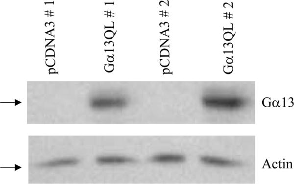
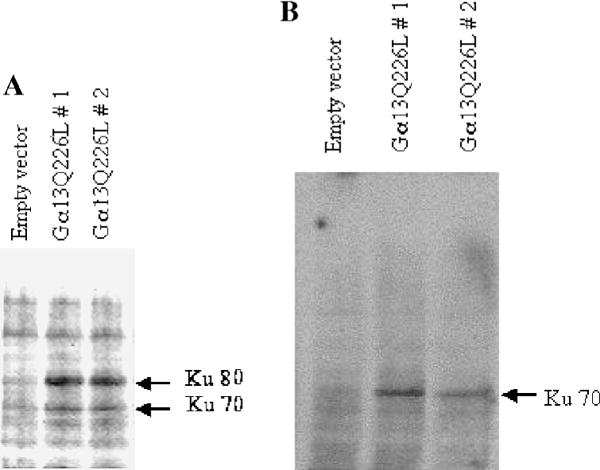
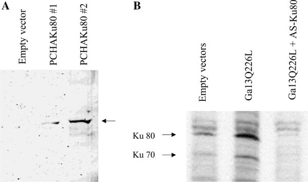
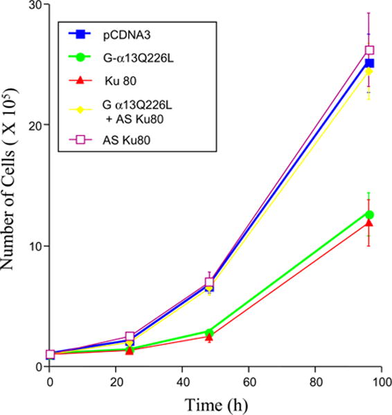
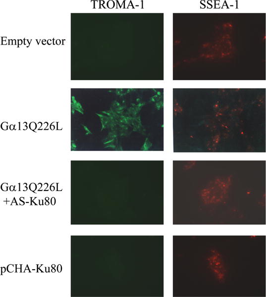
We have shown that a constitutively active Galpha13 (Galpha13Q226L) induces differentiation in P19 embryonic carcinoma cells to an endodermal phenotype. In this report, we demonstrate that Ku, a heterodimer of p80 (Ku80) and p70 (Ku70), is upregulated in P19 cells overexpressing Galpha13Q226L. Ku is the regulatory subunit of the DNA-dependent protein kinase and is primarily involved in DNA repair and recombination. Ku80 also is a somatostatin receptor. We show that while overexpression of Ku80 drastically reduced P19 cell proliferation, it was not sufficient to induce endodermal differentiation. However, coexpression of Galpha13Q226L and an antisense Ku80 abrogated the retarded growth rate and endodermal differentiation observed in cells expressing only Galpha13Q226L. Overexpression of Galpha13Q226L or Ku80 downregulated RNA polymerase I-mediated transcriptional activity and overexpression of antisense Ku80 restored the activity to control level. These results suggest that Ku80 is required for Galpha13-mediated endodermal differentiation in P19 cells.


Figures

Fig. 1
Fig. 1
P19 cells transfected with Gα13Q226L plasmid ectopically express the constitutively active Gα13Q226L protein. Total protein was purified from various stable P19 clones. One hundred micrograms of protein/ lane was separated by SDS–polyacrylamide gel electrophoresis on 10% gels, transferred to nitrocellulose membranes, and probed with primary antibodies to Gα13. To confirm equal loading, an antibody against actin was used to detect actin in the various extracts (bottom panel).
Fig. 2
Fig. 2
Constitutively active Gα13 upregulates Ku (Ku80/Ku70) protein level in P19 cells. An immunoblot analysis was performed taking 100 μg of total protein in each lane of a 7.5% SDS–polyacryl-amide gel. Anti-Ku antiserum (OM) that recognizes the human and murine Ku heterodimer (A) and a monoclonal antibody (N3H10) that recognizes Ku70 (B) were used. Two different clones of Gα13Q226L expressing cells are shown in comparison to the empty vector control. The Ku polypeptides are marked with arrows.
Fig. 3
Fig. 3
Overexpression of Ku80 in P19 cells is achieved by transfection with pCHA-Ku80 and downregulation of Ku80 is induced by transfection of an antisense Ku80, AS-Ku80. (A) P19 cells were stably transfected with PCHA-Ku80 and pCHA. Immunoblot analysis of the cell extracts was performed using an antibody (anti-HA tag) against the hemaglutinnin tag polypeptide expressed from the C-terminal end of the pCHA-Ku80 vector. The position of HA-Ku80 is marked with an arrow. (B) P19 cells were transfected either with PCDNA3 plus pT-Hygr (lane, empty vectors), or PCDNA3 Gα13Q226L plus pT-Hygr (lane, Gα13Q226L), or PCDNA3 Gα13Q226L plus pT-HygrAS-Ku80 (lane, Gα13Q226L + AS-Ku80). Immunoblot analysis of cell extracts was performed using the anti-Ku antiserum (OM). Arrows indicate the positions of Ku80 and Ku70.
Fig. 4
Fig. 4
Antisense Ku80 relieves P19 cells from growth retardation induced by the constitutively active Gα13Q226L. Cells were stably transfected separately with PCDNA3, Gα13Q226L, pCHAKu80, Gα13Q226L plus pT-HygrAS-Ku80, and pT-HygrAS-Ku80. Proliferation curve of P19 clones overexpressing different proteins was obtained. Analyses of multiple clones were performed, and a representative clone for each cell line is presented. Triplicate culture wells for each time point were established at time zero. Error bars represent the standard deviation about the mean values.
Fig. 5
Fig. 5
Coexpression of constitutively active Gα13Q226L and antisense Ku80 (AS-Ku80) inhibits P19 cell differentiation and constitutively active Q226L stimulates differentiation to an endodermal phenotype. Stable clones of cells expressing either the empty vectors (PCDNA3 and pT-Hygr) or Gα13Q226L (PCDNA3Gα13Q226L + pT-Hygr) or Gα13Q226L and AS-Ku80 (PCDNA3Gα13Q226L + pTHygrAS-Ku80) were subjected to indirect immunofluorescence after staining with antibodies specific for an embryonal marker (SSEA-1) or endodermal marker (TROMA-1).

Similar articles

Cited by

References

    1. Dvir A, Peterson SR, Knuth MW, Lu H, Dynan WS. Ku autoantigen is the regulatory component of a template-associated protein kinase that phosphorylates RNA polymerase II. Proc Natl Acad Sci USA. 1992;89:11920–11924. - PMC - PubMed
    1. Mimori T, Hardin JA, Steitz JA. Characterization of the DNA-binding protein antigen Ku recognized by autoantibodies from patients with rheumatic disorders. J Biol Chem. 1986;261:2274–2278. - PubMed
    1. Le Romancer ML, Reyl-Desmars F, Cherifi Y, Pigeon C, Bottari S, Meyer O, Lewin MJM. The 86-kDa subunit of autoantigen Ku is a somatostatin receptor regulating protein phosphatase-2A activity. J Biol Chem. 1994;269:7464–17468. - PubMed
    1. Janssens V, Goris J. Protein phosphatase 2A: a highly regulated family of serine/threonine phosphatases implicated in cell growth and signaling. Biochem J. 2001;353:417–439. - PMC - PubMed
    1. Scheer U, Zentgraf H, pug A. In: The Cell Nucleus II. Busch H, Rothblum L, editors. Academic Press; New York: 1982. pp. 143–176.

Publication types

MeSH terms