. 2004 Sep 13;166(6):889-900.
doi: 10.1083/jcb.200405061. Epub 2004 Sep 7.

Robust cell polarity is a dynamic state established by coupling transport and GTPase signaling

Affiliations

Robust cell polarity is a dynamic state established by coupling transport and GTPase signaling

Roland Wedlich-Soldner et al. J Cell Biol. .

Abstract

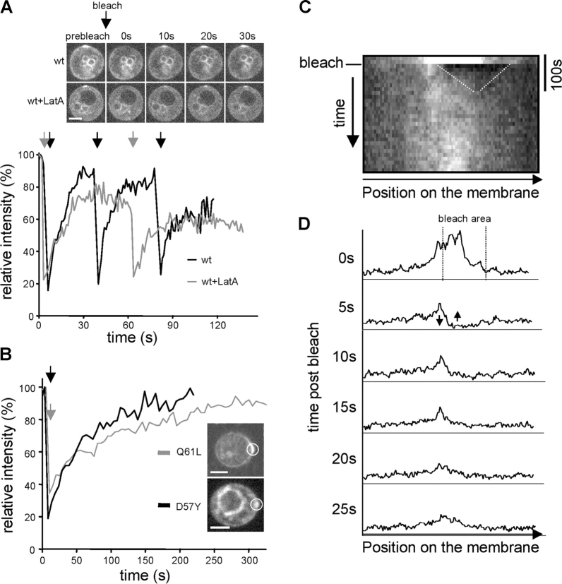
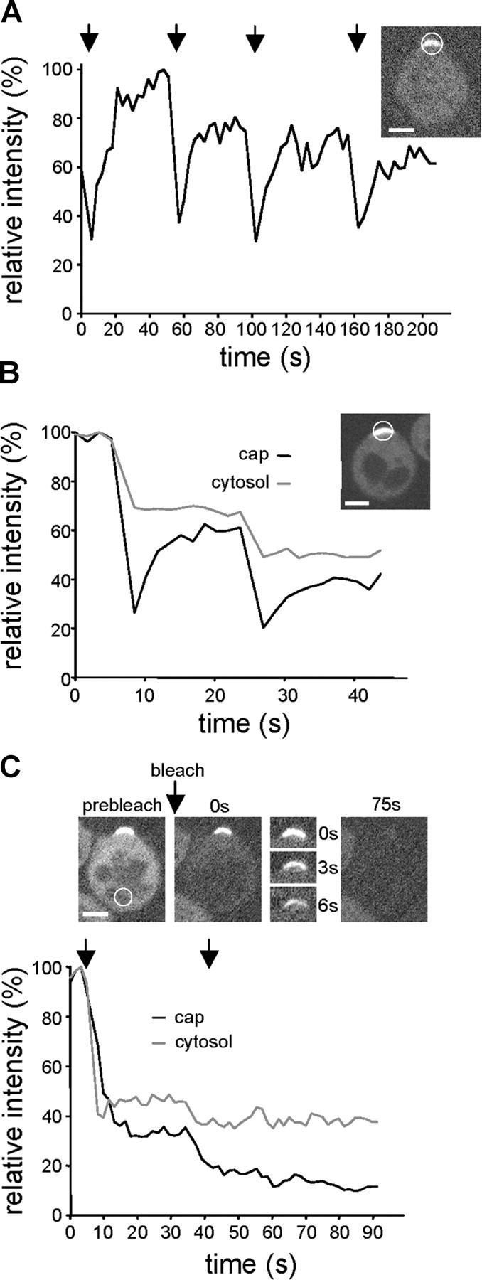
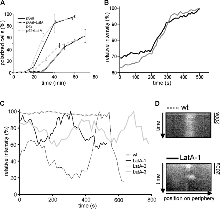
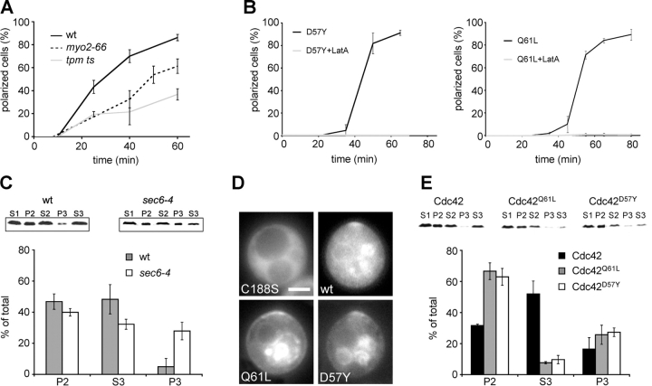
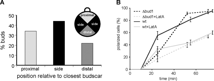
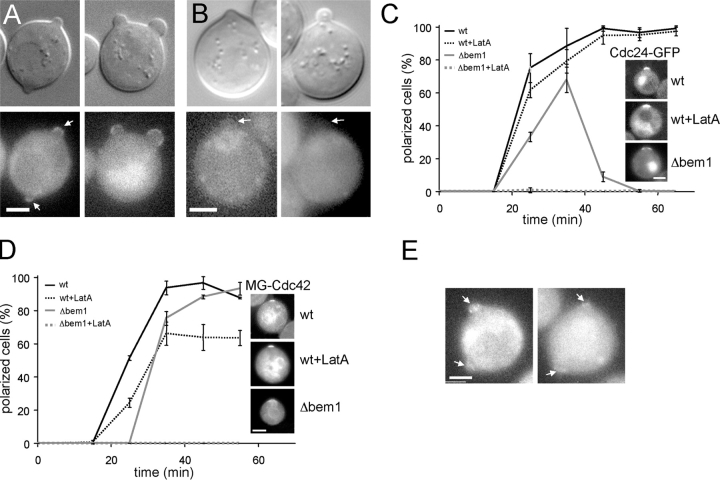
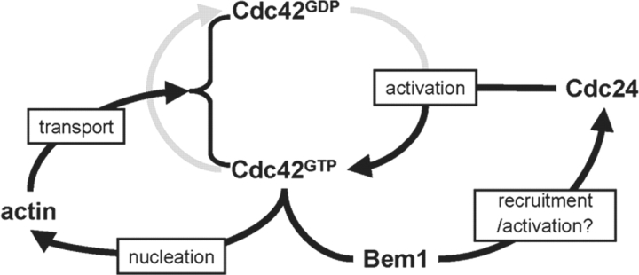
Yeast cells can initiate bud formation at the G1/S transition in a cue-independent manner. Here, we investigate the dynamic nature of the polar cap and the regulation of the GTPase Cdc42 in the establishment of cell polarity. Using analysis of fluorescence recovery after photobleaching, we found that Cdc42 exchanged rapidly between the polar caps and cytosol and that this rapid exchange required its GTPase cycle. A previously proposed positive feedback loop involving actomyosin-based transport of the Cdc42 GTPase is required for the generation of robust cell polarity during bud formation in yeast. Inhibition of actin-based transport resulted in unstable Cdc42 polar caps. Unstable polarity was also observed in mutants lacking Bem1, a protein previously implicated in a feedback loop for Cdc42 activation through a signaling pathway. When Bem1 and actin were both inhibited, polarization completely failed. These results suggest that cell polarity is established through coupling of transport and signaling pathways and maintained actively by balance of flux.


Figures

Figure 1.
Figure 1.
FRAP analysis of MG-Cdc42 polar caps. In all graphs, the time points of bleaching are indicated by arrows and the area of bleaching is indicated by circles. Average intensities are given relative to the prebleach state. Bars, 5 μm. (A) FRAP of MG-Cdc42 caps formed in RLY1948 cells 30 min after release from G1 arrest. Caps formed in the presence (wt+LatA, gray line) or absence (wt, black line) of LatA were bleached multiple times and recovery monitored by time-lapse microscopy at 1 frame/s. Images from representative examples show caps before bleaching and at the indicated times after bleaching. (B) FRAP of MG-Cdc42Q61L (RLY1703) and MG-Cdc42D57Y (RLY1991) caps 35 min after release from G1 arrest. Recovery was monitored as in A with frames taken every 5 s. (C) Chymograph showing recovery of the MG-Cdc42Q61L cap shown in B. The initial recovery by lateral diffusion is indicated by dotted lines. (D) Line scans of the MG-Cdc42Q61L cap analyzed in B and C at the indicated times after bleaching (0 s represents the prebleach state). Arrows at the 5-s time point indicate the equalizing of fluorescence by diffusion seen during the initial recovery period.
Figure 2.
Figure 2.
FRAP analysis of Cdc24-GFP and Bem1-GFP polar caps. The time points of bleaching are indicated by arrows and the area of bleaching is indicated by circles. Average intensities are given relative to the prebleach state. Images were taken every second. Bars, 5 μm. (A) FRAP of a Cdc24-GFP cap in an RLY1891 cell formed 25 min after release of cells from G1 arrest. One representative example of multiple bleaching and recovery is shown. (B) FRAP of a Bem1-GFP cap in an RLY1963 cell formed 25 min after release from G1 arrest. Fluorescence was monitored both in the cap (black line) and the cytosol (gray line). (C) Fluorescence loss in photobleaching of Bem1-GFP in an RLY1963 cell in the cytosol 25 min after release from G1 arrest. After bleaching the cytosol at the opposite side of the Bem1-GFP cap, fluorescence was measured in the cytosol (gray line) and the cap (black line). Images show the whole cell or the cap area at the indicated times after bleaching.
Figure 3.
Figure 3.
Polarization of Cdc42 after release from G1 arrest. (A) Polarization of MG-Cdc42 expressed from the Gal1 promoter (pGal, RLY1948) or the CDC42 promoter (p42, RLY1951) upon release from G1 arrest in the presence or absence of LatA. The percentage of cells with polarized MG-Cdc42 was determined at different time points (given in min) after release. Error bars correspond to SD. (B) Kinetics of MG-Cdc42 cap formation in two representative wild-type (RLY1950) cells determined by time-lapse imaging started at 10 min after release from G1 arrest. Fluorescence intensity in the cap was measured every 20 s. (C) MG-Cdc42 caps monitored in the presence or absence of LatA by time-lapse imaging 20 min after release from G1 arrest. Normalized average intensities in the cap region were measured every 15 s and plotted against time. (D) Chymographs showing the stability of two caps monitored in C.
Figure 4.
Figure 4.
A role for actin-based membrane transport in the polarization of wild-type and mutant Cdc42. (A) Polarization of MG-Cdc42 in strains temperature sensitive for actin-based transport upon release from G1 arrest at 35°C. Polarization was assayed as in Fig. 3 A for strains wt (RLY1948), myo2-66 (RLY1954), and tpm ts (RLY1897). (B) Polarization of MG-Cdc42Q61L (RLY1703) and MG-Cdc42D57Y (RLY1991) upon release from G1 arrest in the presence or absence of LatA. Note that the graphs for the LatA-treated cells run along the x axis. (A and B) Error bars correspond to SD. (C) Membrane fractionation of MG-Cdc42 in wild-type (RLY1948) and sec6-4 (RLY1894) mutant backgrounds. Cell extracts were prepared and separated by differential centrifugation as described in Materials and methods. In brief, cleared cell lysates (S1) were separated into S2 and P2 in a 10,000 g centrifugation step and S2 was further separated into S3 and P3 in a 100,000 g step. A representative example is shown for each strain. Quantification was performed on an Odyssey imager and is represented as an average of three independent experiments. All amounts are normalized to the total present in the lysate. Because loss of material occurred in the pellet fractions P values were calculated from the respective S values. (D) Localization of MG-Cdc42, MG-Cdc42C188S (RLY1952), MG-Cdc42Q61L, and MG-Cdc42D57Y 25 min after release from G1 arrest. Whereas MG-Cdc42C188S was mostly in the cytosol, all prenylated forms of Cdc42 were present on membranes to varying degrees. MG-Cdc42 showed a clear cytosolic pool (note cytosolic fluorescence in top right panel). MG-Cdc42Q61L and MG-Cdc42D57Y were mostly found on internal membranes and the plasma membrane. Bar, 5 μm. (E) Membrane fractionation of different Cdc42 forms. Cell extracts for strains RLY1948, RLY1703, and RLY1991 were prepared and separated by differential centrifugation as described in C. A representative example is shown for each Cdc42 form. Quantification is represented as average of two independent experiments.
Figure 5.
Figure 5.
Cdc42 polarization is independent of bud site selection. (A) Budding pattern of RLY1683 cells. The position of the first bud emerging after release from G1 arrest was scored in relation to the closest bud scar as proximal, distal or to the side (see diagram). (B) Effect of BUD1 deletion on MG-Cdc42 polarization. Wild-type (RLY1948) or Δbud1 (RLY1959) cells were released from G1 arrest and polarization of MG-Cdc42 was scored in the presence or absence of LatA. Error bars correspond to SD.
Figure 6.
Figure 6.
Actin and Bem1 function in parallel during cell polarization. (A) Recruitment of Cdc24-GFP by Cdc42Q61L. HA-tagged Cdc42Q61L was expressed from the Gal1-promoter in G1-arrested cells that also expressed Cdc24-GFP (RLY1989). Representative DIC and fluorescence images of cells with two buds, one of which must be ectopic. (B) Cdc24-GFP is not recruited to buds of Δbem1 cells expressing HA-Cdc42Q61L. Representative DIC and fluorescence images of cells with small and medium buds are shown. (A and B) Buds are indicated with arrows. (C) Kinetics of polarization of Cdc24-GFP expressed from the CDC24 promoter in wild-type (RLY1891) or Δbem1 (RLY1961) cells released from G1 arrest. Images show typical caps formed at 35 min after release. Note that the graph for the LatA-treated Δbem1 cells runs along the x axis. (D) Kinetics of polarization of MG-Cdc42 expressed from the CDC42 promoter in wild-type (RLY1950) or Δbem1 (RLY1965) cells released from G1 arrest. Images show typical caps formed at 35 min after release. Note that the graph for the LatA-treated Δbem1 cells runs along the x axis. Error bars correspond to SD. (E) Localization of Cdc24-GFP to multiple caps in Δbem1 cells 25 min after release from G1 arrest. Caps are indicated with arrows. Bars, 5 μm.
Figure 7.
Figure 7.
A schematic diagram depicting the two positive feedback loops (in dark lines) that control the generation of cue- independent cell polarity in yeast. The actin-dependent feedback loop results in accumulation of Cdc42 in both nucleotide-bound forms on the plasma membrane, whereas the Bem1-dependent feedback loop results in recruitment and/or activation of the GEF Cdc24 to the site of Cdc42 accumulation. Coupling of these feedback loops is required for the generation of robust cell polarity.

References

    1. Amon, A., S. Irniger, and K. Nasmyth. 1994. Closing the cell cycle circle in yeast: G2 cyclin proteolysis initiated at mitosis persists until the activation of G1 cyclins in the next cycle. Cell. 77:1037–1050. - PubMed
    1. Ayscough, K.R., J. Stryker, N. Pokala, M. Sanders, P. Crews, and D.G. Drubin. 1997. High rates of actin filament turnover in budding yeast and roles for actin in establishment and maintenance of cell polarity revealed using the actin inhibitor latrunculin-A. J. Cell Biol. 137:399–416. - PMC - PubMed
    1. Bagnat, M., and K. Simons. 2002. Cell surface polarization during yeast mating. Proc. Natl. Acad. Sci. USA. 99:14183–14188. - PMC - PubMed
    1. Becskei, A., B. Seraphin, and L. Serrano. 2001. Positive feedback in eukaryotic gene networks: cell differentiation by graded to binary response conversion. EMBO J. 20:2528–2535. - PMC - PubMed
    1. Blumer, K.J., and J.A. Cooper. 2003. Go ahead, break my symmetry! Nat. Cell Biol. 5:1048–1049. - PMC - PubMed

Publication types

MeSH terms

Substances