. 2004 Sep 6;200(5):647-57.
doi: 10.1084/jem.20040646.

Dormancy phenotype displayed by extracellular Mycobacterium tuberculosis within artificial granulomas in mice

Affiliations

Dormancy phenotype displayed by extracellular Mycobacterium tuberculosis within artificial granulomas in mice

Petros C Karakousis et al. J Exp Med. .

Abstract

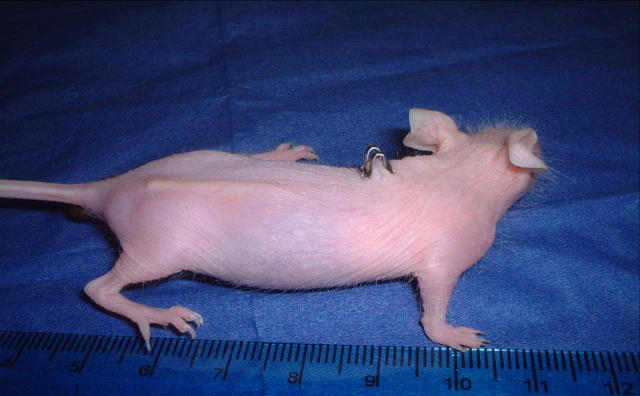
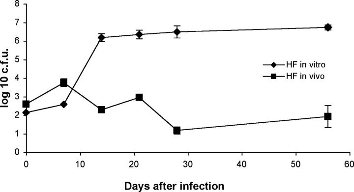
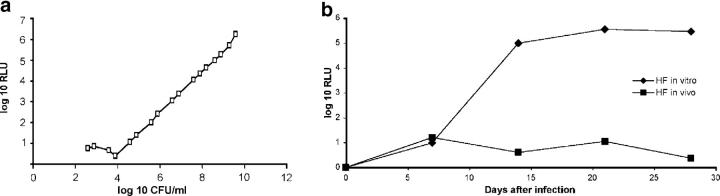
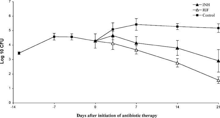
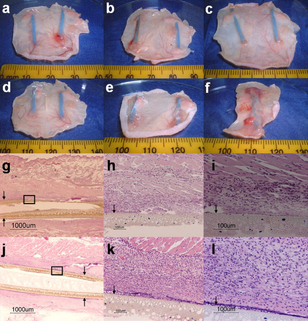
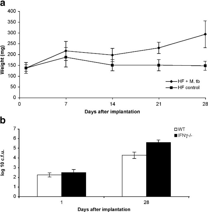
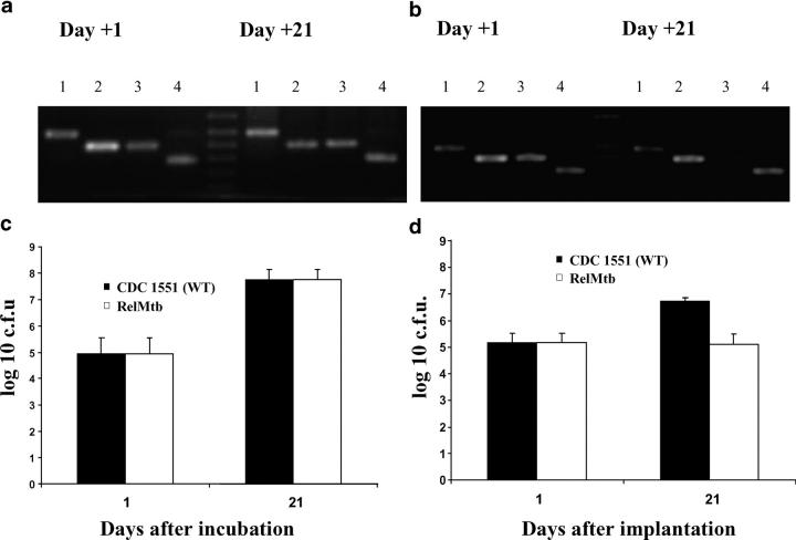
Mycobacterium tuberculosis residing within pulmonary granulomas and cavities represents an important reservoir of persistent organisms during human latent tuberculosis infection. We present a novel in vivo model of tuberculosis involving the encapsulation of bacilli in semidiffusible hollow fibers that are implanted subcutaneously into mice. Granulomatous lesions develop around these hollow fibers, and in this microenvironment, the organisms demonstrate an altered physiologic state characterized by stationary-state colony-forming unit counts and decreased metabolic activity. Moreover, these organisms show an antimicrobial susceptibility pattern similar to persistent bacilli in current models of tuberculosis chemotherapy in that they are more susceptible to the sterilizing drug, rifampin, than to the bactericidal drug isoniazid. We used this model of extracellular persistence within host granulomas to study both gene expression patterns and mutant survival patterns. Our results demonstrate induction of dosR (Rv3133c) and 20 other members of the DosR regulon believed to mediate the transition into dormancy, and that rel(Mtb) is required for Mycobacterium tuberculosis survival during extracellular persistence within host granulomas. Interestingly, the dormancy phenotype of extracellular M. tuberculosis within host granulomas appears to be immune mediated and interferon-gamma dependent.


Figures

Figure 1.
Figure 1.
SKH1 mouse with subcutaneously implanted hollow fiber containing M. tuberculosis.
Figure 2.
Figure 2.
Reduced growth of bacilli within hollow fibers in vivo. CFU counts per fiber of hollow fiber–encapsulated M. tuberculosis implanted into mice (HF in vivo) are compared with those of hollow fiber–encapsulated M. tuberculosis incubated in vitro (HF in vitro).
Figure 3.
Figure 3.
Hollow fiber–encapsulated bacilli in vivo remain viable. As a control, in vitro–grown cultures of M. tuberculosis H37Rv-lux were treated with 70% ethanol for 3 h to promote bacillary death (a). Live bacilli exhibit green fluorescence, whereas dead bacilli fluoresce red. Approximately half of all in vivo hollow fiber–encapsulated organisms on days 21 (b) and 28 (c) after fiber implantation were determined to be viable based on their staining properties.
Figure 4.
Figure 4.
Reduced metabolic activity of encapsulated bacilli in vivo. (a) Relationship of relative light units (RLUs) to CFUs in mid-log phase M. tuberculosis H37Rv-lux grown in vitro. (b) Luciferase activity of hollow fiber–encapsulated M. tuberculosis implanted into mice (HF in vivo) versus hollow fiber–encapsulated M. tuberculosis incubated in vitro (HF in vitro).
Figure 5.
Figure 5.
Hollow fiber–encapsulated bacilli are more susceptible to rifampin than to isoniazid. The activities of isoniazid 0.05% in the diet (INH) and rifampin 0.02% in the diet (RIF) against hollow fiber–encapsulated bacilli in vivo are compared with no treatment (Control).
Figure 6.
Figure 6.
Formation of granuloma-like lesions surrounding M. tuberculosis–containing hollow fibers. Gross skin lesions surrounding hollow fibers containing liquid broth alone at days 1 (a), 14 (b), and 28 (c), and those containing M. tuberculosis H37Rv-lux at days 1 (d), 14 (e), and 28 (f) after hollow fiber implantation. Histopathology of tissues surrounding hollow fibers containing liquid broth (g–i) and those containing M. tuberculosis H37Rv-lux (j–l) 28 d after hollow fiber implantation (hematoxylin-eosin stain). Arrows indicate hollow fiber membrane.
Figure 7.
Figure 7.
Containment of intrafiber bacillary growth in vivo is immune mediated and IFNγ dependent. Mice implanted with hollow fibers containing M. tuberculosis (HF + M. tb) developed enlarged spleens as measured by mean spleen weight (y axis) as early as 14 d after implantation as compared with mice implanted with fibers containing media (HF control; a). Wild-type BALB-C/J (WT) mice are able to contain the growth of hollow fiber–encapsulated M. tuberculosis to a greater extent than isogenic IFNγ-deficient (IFNγ−/−) mice 28 d after hollow fiber implantation (b).
Figure 8.
Figure 8.
Absence of rel Mtb-deficient mutant by PCR from a pool of mutants after 21 d of cultivation within mouse granulomas. PCR amplification of the Tn insertion junction (see Online Supplemental Material) reveals presence of the rel Mtb::Tn mutant in both input (day 1) and output (day 21) pools in hollow fibers incubated in vitro (a), but absence of the mutant in the output pool (day 21) in mouse-implanted hollow fibers (b), suggesting reduced survival of this mutant in vivo. 1, Rv1347 (hypothetical transcriptional regulator); 2, Rv0250 (miscellaneous oxidoreductase); 3, Rv2583c (rel Mtb); 4, Rv1069 (pra). Hollow fiber–encapsulated wild-type M. tuberculosis CDC 1551 (WT) and rel Mtb::Tn mutant (RelMtb) grow equally when incubated in hollow fibers in vitro (c), but the latter strain demonstrates significantly reduced survival when hollow fibers are implanted into mice (d).

References

    1. Cegielski, J.P., D.P. Chin, M.A. Espinal, T.R. Frieden, R. Rodriquez Cruz, E.A. Talbot, D.E. Weil, R. Zaleskis, and M.C. Raviglione. 2002. The global tuberculosis situation. Progress and problems in the 20th century, prospects for the 21st century. Infect. Dis. Clin. North Am. 16:1–58. - PubMed
    1. Flynn, J.L., and J. Chan. 2001. Tuberculosis: latency and reactivation. Infect. Immun. 69:4195–4201. - PMC - PubMed
    1. Kaplan, G., F.A. Post, A.L. Moreira, H. Wainwright, B.N. Kreiswirth, M. Tanverdi, B. Mathema, S.V. Ramaswamy, G. Walther, L.M. Steyn, et al. 2003. Mycobacterium tuberculosis growth at the cavity surface: a microenvironment with failed immunity. Infect. Immun. 71:7099–7108. - PMC - PubMed
    1. Grosset, J. 2003. Mycobacterium tuberculosis in the extracellular compartment: an underestimated adversary. Antimicrob. Agents Chemother. 47:833–836. - PMC - PubMed
    1. Talaat, A.M., R. Lyons, S.T. Howard, and S.A. Johnston. 2004. The temporal expression profile of Mycobacterium tuberculosis infection in mice. Proc. Natl. Acad. Sci. USA. 101:4602–4607. - PMC - PubMed

Publication types

MeSH terms