. 2004 Sep 6;200(5):659-69.
doi: 10.1084/jem.20040789.

Activation of PI3K is indispensable for interleukin 7-mediated viability, proliferation, glucose use, and growth of T cell acute lymphoblastic leukemia cells

Affiliations

Activation of PI3K is indispensable for interleukin 7-mediated viability, proliferation, glucose use, and growth of T cell acute lymphoblastic leukemia cells

Joao T Barata et al. J Exp Med. .

Abstract

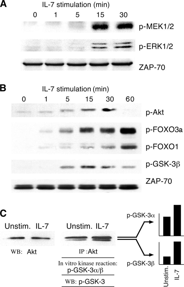
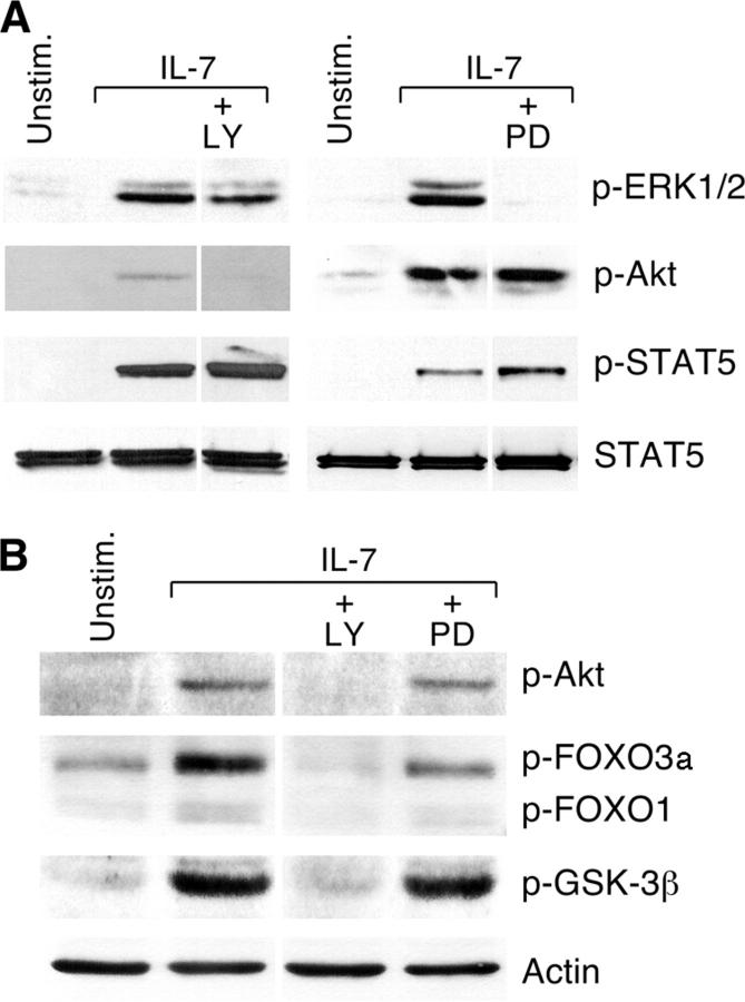
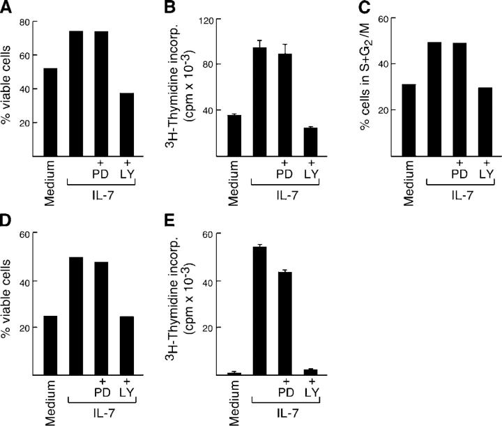
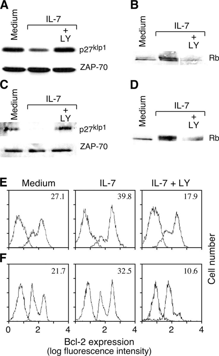
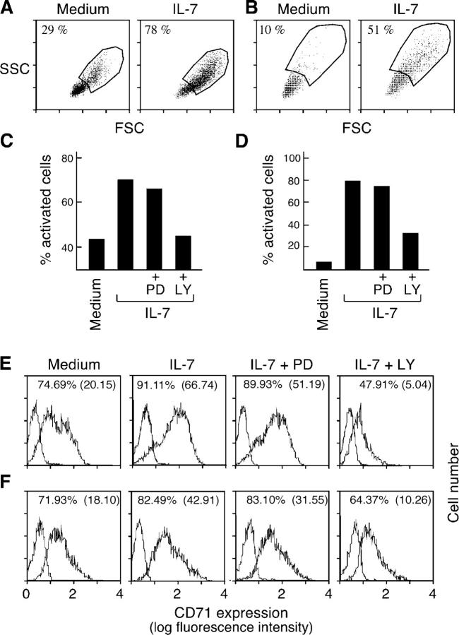
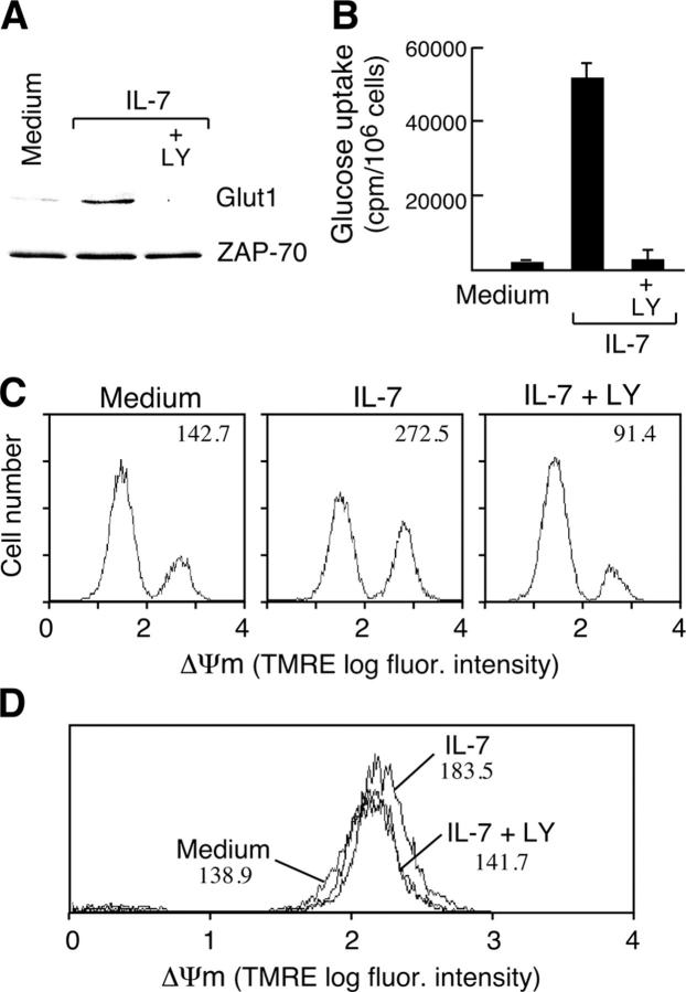
Interleukin (IL)-7 is essential for normal T cell development. Previously, we have shown that IL-7 increases viability and proliferation of T cell acute lymphoblastic leukemia (T-ALL) cells by up-regulating Bcl-2 and down-regulating the cyclin-dependent kinase inhibitor p27kip1. Here, we examined the signaling pathways via which IL-7 mediates these effects. We investigated mitogen-activated protein kinase (MEK)-extracellular signal-regulated kinase (Erk) and phosphatidylinositol-3-kinase (PI3K)-Akt (protein kinase B) pathways, which have active roles in T cell expansion and have been implicated in tumorigenesis. IL-7 induced activation of the MEK-Erk pathway in T-ALL cells; however, inhibition of the MEK-Erk pathway by the use of the cell-permeable inhibitor PD98059, did not affect IL-7-mediated viability or cell cycle progression of leukemic cells. IL-7 induced PI3K-dependent phosphorylation of Akt and its downstream targets GSK-3, FOXO1, and FOXO3a. PI3K activation was mandatory for IL-7-mediated Bcl-2 up-regulation, p27kip1 down-regulation, Rb hyperphosphorylation, and consequent viability and cell cycle progression of T-ALL cells. PI3K signaling was also required for cell size increase, up-regulation of CD71, expression of the glucose transporter Glut1, uptake of glucose, and maintenance of mitochondrial integrity. Our results implicate PI3K as a major effector of IL-7-induced viability, metabolic activation, growth and proliferation of T-ALL cells, and suggest that PI3K and its downstream effectors may represent molecular targets for therapeutic intervention in T-ALL.


Figures

Figure 1.
Figure 1.
IL-7 activates MEK–Erk and PI3K–Akt pathways in T-ALL. IL-7–deprived TAIL7 cells were stimulated with IL-7 for the indicated periods (A and B). Cell lysates were resolved with 10% SDS-PAGE and immunoblotted with the indicated antibodies. Results representative of three independent experiments are shown. Levels of phosphorylated MEK1/2 and Erk1/2 were detected with antisera that selectively recognize the activated forms of the following kinases: Ser217/Ser221-phosphorylated MEK1/2 (P-MEK1/2) and Thr202/Tyr204-dual-phosphorylated Erk1/2 (P-Erk1/2). Levels of phosphorylated Akt and GSK-3 were analyzed with antibodies that specifically recognize Ser473-phosphorylated Akt (P-Akt) and Ser9-phosphorylated GSK-3β (P-GSK-3β), respectively. Levels of phosphorylated FOXO1/FOXO3a were detected with antiserum that reacts with Thr24-phosphorylated FOXO1 (P-AFOXO1) and Thr32-phosphorylated FOXO3a (P- FOXO3a). Akt and Erk1/2 protein levels were assessed with specific antibodies and remained unchanged (not depicted). Blots were reprobed with an anti–ZAP-70 antibody to confirm even protein loading. (C) IL-7 activates Akt and induces in vitro phosphorylation of GSK-3 by Akt. IL-7–deprived TAIL7 cells were stimulated with IL-7 for 15 min. To compare Akt enzymatic activity in unstimulated (Unst.) versus IL-7–stimulated cells (IL-7), cell lysates were immunoprecipitated with agarose-conjugated anti-Akt antibody and in vitro kinase reactions were performed using crosstide-GSK-3α/β as exogenous substrate. Reactions were analyzed by 12% SDS-PAGE, transferred to nitrocellulose membrane, and GSK-3 phosphorylation was detected by immunoblotting with anti–phospho-GSK-3α/β (Ser21/Ser9) antibody. Even loading was confirmed with an anti-Akt antibody. Relative quantification of phosphorylated GSK-3α and GSK-3β bands was performed by densitometry analysis. Results were normalized in relation to the loading control (Akt) and expressed as relative units. IL-7 induced a 1.63-fold increase in GSK-3α and a 3.07-fold increase in GSK-3β Akt-mediated phosphorylation. Results are representative of two independent experiments.
Figure 2.
Figure 2.
IL-7 induces PI3K-dependent phosphorylation of Akt, GSK-3, FOXO1, and FOXO3a, and MEK-dependent phosphorylation of Erk1/2 in T-ALL cells. IL-7–deprived TAIL7 cells were pretreated with 10 μM LY294002 (LY) or 10 μM PD098059 (PD) for 2 h, and then stimulated with IL-7 for 15 min. (A) Western blot analysis was performed with P-Erk1/2, P-Akt antibodies (see legend to Fig. 1), and an antibody specific for Tyr694/Tyr699-phosphorylated-STAT5A/B (P-STAT5) to confirm that LY294002 and PD98059 were specific inhibitors of the PI3K–Akt and MEK–Erk pathway, respectively. (B) GSK-3β, FOXO1, and FOXO3a phosphorylation is dependent on PI3K activity. Western blot analysis was performed with P-Akt, P-GSK3β, and P-FOXO1/FOXO3a antibodies. Anti-STAT5 (A) and actin (B) antibodies were used to confirm equal loading. Representative results from three independent experiments are shown.
Figure 3.
Figure 3.
PI3K but not MEK is critical for IL-7–mediated viability and cell cycle progression of T-ALL cells. (A–C) TAIL7 cells were cultured for 96 h with 10 ng/ml IL-7, either alone or in the presence of 10 μM PD098059 (IL-7+PD) or 10 μM LY294002 (IL-7+LY). (A) TAIL7 cells were stained with annexin V–FITC plus propidium iodide and viability was determined by flow cytometry analysis. (B) Proliferation was determined by assessment of [3H]thymidine incorporation. (C) Percentage of cells at S+G2/M phases of the cell cycle was determined by propidium iodide staining followed by flow cytometry analysis. (D and E) Primary T-ALL cells were obtained as described in Materials and Methods, and cultured under the indicated conditions. Viability at 96 h (D) and proliferation at 72 h (E) of culture were assessed as described for TAIL7 cells. Results are representative of three to six independent experiments with TAIL7 cell line and two independent experiments with all five primary T-ALL samples. Results from remaining primary T-ALL samples are shown in Fig. S1.
Figure 4.
Figure 4.
IL-7 mediates p27kip1 down-regulation, Rb hyperphosphorylation, and Bcl-2 up-regulation via activation of PI3K in T-ALL cells. TAIL7 cells (A and B) or primary T-ALL cells (C and D) were cultured for 96 or 72 h, respectively, under the indicated conditions. (A and C) Cell lysates were resolved by 10% SDS-PAGE and immunoblotted with anti-p27kip1 antibody. Membranes were stripped and reprobed with ZAP-70 to confirm equal loading. (B and D) Lysates from the same samples were analyzed by 6% SDS-PAGE and immunoblotted with an Rb-specific antibody. The hyperphosphorylated form of Rb corresponds to the band with the higher apparent molecular weight. Blasts from T-ALL number 3 were used in this experiment. (E) Bcl-2 protein levels at 96 h of culture were assessed by flow cytometry after intracellular staining of TAIL7 cells with FITC-conjugated anti–Bcl-2 antibody. (F) Expression of Bcl-2 in primary T-ALL cells was assessed at 72 h of culture. Results are representative of four different patient samples analyzed. Specific MIF, as described in Materials and Methods, is indicated in each histogram. Results were similar in six independent experiments.
Figure 5.
Figure 5.
PI3K is critical for IL-7–mediated cell growth and activation of T-ALL cells. TAIL7 (A) and primary T-ALL (B) cells were cultured for 72 h in medium alone or with 10 ng/ml IL-7, and then analyzed by flow cytometry for changes in cell size (determined by FSC) in the live cell population. Percentage of “activated” cells was calculated by defining a threshold gate that excluded the bulk, small-sized population of medium-cultured cells. Results from one representative patient of five tested are shown in B. Results shown in A are similar to those found in numerous independent experiments. TAIL7 cells (C) and primary T-ALL cells (D) were cultured for 96 h under the indicated conditions, and the percentage of activated cells was calculated. TAIL7 cells cultured for 96 h (E) and primary T-ALL cells cultured for 72 h (F) under the indicated conditions were stained with anti-CD71 antibody. Results were expressed as the percentage of positive cells and as the specific MIF (in brackets). Results from primary T-ALL cells and the TAIL7 cell line were similar in three independent experiments.
Figure 6.
Figure 6.
IL-7 regulates Glut1 expression and mitochondrial homeostasis via PI3K activation. TAIL7 cells were cultured for 96 h under the indicated conditions. (A) Cell lysates were resolved with 10% SDS-PAGE and immunoblotted with anti-Glut1 antibody. Anti–ZAP-70 antibody was used in the same membrane to ascertain even protein loading. (B) IL-7 promotes glucose uptake in a PI3K-dependent manner. TAIL7 cells were cultured as indicated for 96 h. Cells were then assayed for glucose uptake as described in Materials and Methods. (C) Mitochondrial membrane potential (Δψm) was assessed at 96 h of culture by staining TAIL7 cells with the potentiometric dye TMRE and analyzing the whole population by flow cytometry. TMRE intensity reflects the Δψm. Results are expressed as MIF. (D) Viable cells were identified and selected by flow cytometry by gating on the live cell population, as determined by FSC × SSC and/or annexin V–FITC staining. Δψm of this population was determined using TMRE. Results are representative of three independent experiments.

Similar articles

Cited by

References

    1. von Freeden-Jeffry, U., N. Solvason, M. Howard, and R. Murray. 1997. The earliest T lineage-committed cells depend on IL-7 for Bcl-2 expression and normal cell cycle progression. Immunity. 7:147–154. - PubMed
    1. Dibirdik, I., M.C. Langlie, J.A. Ledbetter, L. Tuel-Ahlgren, V. Obuz, K.G. Waddick, K. Gajl-Peczalska, G.L. Schieven, and F.M. Uckun. 1991. Engagement of interleukin-7 receptor stimulates tyrosine phosphorylation, phosphoinositide turnover, and clonal proliferation of human T-lineage acute lymphoblastic leukemia cells. Blood. 78:564–570. - PubMed
    1. Karawajew, L., V. Ruppert, C. Wuchter, A. Kosser, M. Schrappe, B. Dorken, and W.D. Ludwig. 2000. Inhibition of in vitro spontaneous apoptosis by IL-7 correlates with bcl-2 up-regulation, cortical/mature immunophenotype, and better early cytoreduction of childhood T-cell acute lymphoblastic leukemia. Blood. 96:297–306. - PubMed
    1. Funk, P.E., R.P. Stephan, and P.L. Witte. 1995. Vascular cell adhesion molecule 1-positive reticular cells express interleukin-7 and stem cell factor in the bone marrow. Blood. 86:2661–2671. - PubMed
    1. Oosterwegel, M.A., M.C. Haks, U. Jeffry, R. Murray, and A.M. Kruisbeek. 1997. Induction of TCR gene rearrangements in uncommitted stem cells by a subset of IL-7 producing, MHC class-II-expressing thymic stromal cells. Immunity. 6:351–360. - PubMed

Publication types

MeSH terms

Substances