Review
. 2004 Sep;68(3):373-402, table of contents.
doi: 10.1128/MMBR.68.3.373-402.2004.

Binary bacterial toxins: biochemistry, biology, and applications of common Clostridium and Bacillus proteins

Affiliations
Review

Binary bacterial toxins: biochemistry, biology, and applications of common Clostridium and Bacillus proteins

Holger Barth et al. Microbiol Mol Biol Rev. 2004 Sep.

Abstract

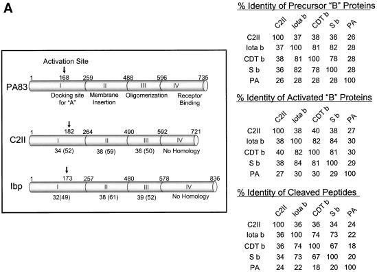
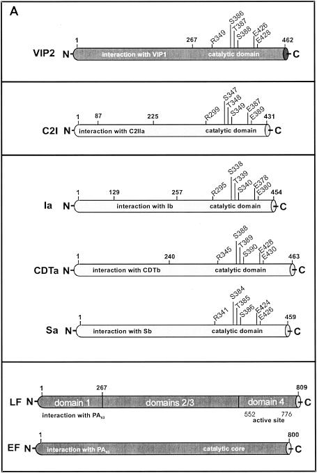
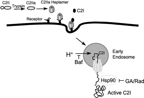
Certain pathogenic species of Bacillus and Clostridium have developed unique methods for intoxicating cells that employ the classic enzymatic "A-B" paradigm for protein toxins. The binary toxins produced by B. anthracis, B. cereus, C. botulinum, C. difficile, C. perfringens, and C. spiroforme consist of components not physically associated in solution that are linked to various diseases in humans, animals, or insects. The "B" components are synthesized as precursors that are subsequently activated by serine-type proteases on the targeted cell surface and/or in solution. Following release of a 20-kDa N-terminal peptide, the activated "B" components form homoheptameric rings that subsequently dock with an "A" component(s) on the cell surface. By following an acidified endosomal route and translocation into the cytosol, "A" molecules disable a cell (and host organism) via disruption of the actin cytoskeleton, increasing intracellular levels of cyclic AMP, or inactivation of signaling pathways linked to mitogen-activated protein kinase kinases. Recently, B. anthracis has gleaned much notoriety as a biowarfare/bioterrorism agent, and of primary interest has been the edema and lethal toxins, their role in anthrax, as well as the development of efficacious vaccines and therapeutics targeting these virulence factors and ultimately B. anthracis. This review comprehensively surveys the literature and discusses the similarities, as well as distinct differences, between each Clostridium and Bacillus binary toxin in terms of their biochemistry, biology, genetics, structure, and applications in science and medicine. The information may foster future studies that aid novel vaccine and drug development, as well as a better understanding of a conserved intoxication process utilized by various gram-positive, spore-forming bacteria.


Figures

FIG. 1.
FIG. 1.
Basic mechanisms of cell intoxication employed by Clostridium and Bacillus binary toxins. Cell-binding “B” precursors are first activated by proteolytic cleavage in solution or on the cell surface. The furin-based, cell-associated cleavage of B. anthracis PA83 is unique, since none of the other “B” precursors are activated after binding to a cell. Activated “B” components interact with a specific cell surface receptor(s) as either preformed, ring-shaped homoheptamers or monomers that subsequently form heptamers. An enzymatic “A” component(s) docks with the cell-bound “B” heptamer, and the receptor-holotoxin complex is then taken up via receptor-mediated endocytosis into early endosomes, which become acidified by vacuolar-type ATPases. An acidic environment is essential for translocating an “A” component(s) into the cytosol, since this induces a conformational change and subsequent insertion of the “B” heptamer into an endosomal membrane to form a channel. Although not clearly defined, it is likely that an “A” component(s) is transported into the cytosol through the “B” heptamer-induced channel.
FIG. 2.
FIG. 2.
The cell-binding “B” proteins of Clostridium and Bacillus binary toxins are activated by serine-type proteases, share varying sequence homology, and form heptameric ring-like structures. (A) Proteolytic cleavage sites, domain functions, and amino acid lengths of PA83, C2II, and Ibp precursor molecules are shown on the left. Below C2II and Ibp are the percent identities (and in parentheses are the percent homologies) for the amino acid sequences from each domain relative to PA83. Percent sequence identities of the “B” precursors (top), activated “B” proteins (middle), and N-terminal peptides released after proteolysis of “B” precursors (bottom) are shown on the right. Sequences were found in either the DNA Data Bank of Japan (DDBJ) with accession number D88982 (C2II) or GenBank with accession numbers M22589 (PA), I40862 (Ib), X97969 (Sb), and L76081 (CDTb). Modified from reference with permission. (B) C2IIa heptamers on lipid bilayers as detected by electron microscopy. Modified from reference with permission.
FIG. 2.
FIG. 2.
The cell-binding “B” proteins of Clostridium and Bacillus binary toxins are activated by serine-type proteases, share varying sequence homology, and form heptameric ring-like structures. (A) Proteolytic cleavage sites, domain functions, and amino acid lengths of PA83, C2II, and Ibp precursor molecules are shown on the left. Below C2II and Ibp are the percent identities (and in parentheses are the percent homologies) for the amino acid sequences from each domain relative to PA83. Percent sequence identities of the “B” precursors (top), activated “B” proteins (middle), and N-terminal peptides released after proteolysis of “B” precursors (bottom) are shown on the right. Sequences were found in either the DNA Data Bank of Japan (DDBJ) with accession number D88982 (C2II) or GenBank with accession numbers M22589 (PA), I40862 (Ib), X97969 (Sb), and L76081 (CDTb). Modified from reference with permission. (B) C2IIa heptamers on lipid bilayers as detected by electron microscopy. Modified from reference with permission.
FIG. 3.
FIG. 3.
(A) Enzymatic “A” proteins of Clostridium and Bacillus binary toxins with known catalytic sites and docking domains for “B” heptamers. (B) ADP-ribosylation of G-actin at R177 by C2I, according to the B. cereus VIP2 model as proposed by Han et al. (171). In the left-hand panel, the hydrophobic cleft of the C2I catalytic domain is depicted with bound NAD. Amino acids E387 to E389 stabilize an intermediate state before nucleophilic attack on G-actin R177, thus yielding mono-ADP-ribosylated G-actin and nicotinamide (right-hand panel). The same mechanism is utilized by other ADP-ribosylating toxins (CDT, CST, and ι) that modify G-actin. Panel B modified from reference with permission.
FIG. 3.
FIG. 3.
(A) Enzymatic “A” proteins of Clostridium and Bacillus binary toxins with known catalytic sites and docking domains for “B” heptamers. (B) ADP-ribosylation of G-actin at R177 by C2I, according to the B. cereus VIP2 model as proposed by Han et al. (171). In the left-hand panel, the hydrophobic cleft of the C2I catalytic domain is depicted with bound NAD. Amino acids E387 to E389 stabilize an intermediate state before nucleophilic attack on G-actin R177, thus yielding mono-ADP-ribosylated G-actin and nicotinamide (right-hand panel). The same mechanism is utilized by other ADP-ribosylating toxins (CDT, CST, and ι) that modify G-actin. Panel B modified from reference with permission.
FIG. 4.
FIG. 4.
Depiction of C2 intoxication via Hsp90, a process that is required for entry of C2I into the cytosol from acidified endosomes (179). A similar pathway is also used by ι toxin and CDT for intoxicating cells (178a). Translocation of C2I, CDTa, or Ia from the endosome requires an acidic pH, a process blocked by bafilomycin A (Baf) via specific inhibition of vacuolar-type ATPases located in the endosomal membrane. C2I partially unfolds during translocation across the endosomal membrane via Hsp90, a process specifically targeted by geldanamycin (GA) or radicicol (Rad), which results in trapping of C2I within the endosome.

Similar articles

Cited by

References

    1. Abrami, L., S. Liu, P. Cosson, S. H. Leppla, and F. G. van der Goot. 2003. Anthrax toxin triggers endocytosis of its receptor via a lipid raft-mediated clathrin-dependent process. J. Cell Biol. 160:3321-3328. - PMC - PubMed
    1. Acheson, D. W. K., and G. T. Keusch. 1999. The family of Shiga toxins, p. 229-242. In J. E. Alouf and J. H. Freer (ed.), The comprehensive sourcebook of bacterial protein toxins. Academic Press, Inc., New York, N.Y.
    1. Agrawal, A., J. Lingappa, S. H. Leppla, S. Agrawal, A. Jabbar, C. Quinn, and B. Pulendran. 2003. Impairment of dendritic cells and adaptive immunity by anthrax lethal toxin. Nature 424:329-334. - PubMed
    1. Ahuja, N., P. Kumar, S. Alam, M. Gupta, and R. Bhatnagar. 2003. Deletion mutants of protective antigen that inhibit anthrax toxin both in vitro and in vivo. Biochem. Biophys. Res. Commun. 307:446-450. - PubMed
    1. Ahuja, N., P. Kumar, and R. Bhatnagar. 2001. Hydrophobic residues Phe552, Phe554, Ile562, Leu566, and Ile574 are required for oligomerization of anthrax protective antigen. Biochem. Biophys. Res. Commun. 287:542-549. - PubMed

Publication types

LinkOut - more resources