Review
. 2004 Sep;68(3):453-73, table of contents.
doi: 10.1128/MMBR.68.3.453-473.2004.

CO-sensing mechanisms

Affiliations
Review

CO-sensing mechanisms

Gary P Roberts et al. Microbiol Mol Biol Rev. 2004 Sep.

Abstract

Carbon monoxide (CO) has long been known to have dramatic physiological effects on organisms ranging from bacteria to humans, but recently there have a number of suggestions that organisms might have specific sensors for CO. This article reviews the current evidence for a variety of proteins with demonstrated or potential CO-sensing ability. Particular emphasis is placed on the molecular description of CooA, a heme-containing CO sensor from Rhodospirillum rubrum, since its biological role as a CO sensor is clear and we have substantial insight into the basis of its sensing ability.


Figures
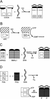
FIG. 1.
FIG. 1.
The general behaviors of CooA, sGC, NPAS, and FixL in response to their effectors. In all cases, the relevant heme pockets are depicted as boxes. (A) In the absence of CO, the CooA homodimer has its DNA-binding surfaces (shaded black) buried away from the solvent. On CO binding, there is a significant rearrangement of a portion of each monomer that allows the DNA-binding surfaces to bind specific DNA sequences. The DNA-bound CooA then interacts with RNA polymerase to allow gene transcription. (B) sGC exists as a heterodimer that is inactive without NO. NO binding to the heme displaces an endogenous histidine ligand (−H), which triggers a conformational change. The active conformation of sGC synthesizes cGMP, an important signal molecule. (C) NPAS2 is only recently described and poorly understood. In the presence of small molecules bound to the heme, the protein exists as an inactive monomer, but the absence of such small molecules leads to the formation of an active heterodimer with another protein, BMAL1, which is able to bind DNA and activate transcription. (D) FixL acts as a homodimer that is inactive when O2 is bound to the hemes. In the absence of O2, FixL undergoes a conformational change that causes autophosphorylation and subsequent transfer of that phosphate to FixJ. Phosphorylated FixJ binds DNA and activates transcription of anaerobically expressed genes.
FIG. 2.
FIG. 2.
Current model for the behavior of the heme vicinity of FixL in response to O2 binding. (A) Active ferrous FixL (Protein Data Bank no. 1LSW), where the heme is depicted at the center, with the Fe atom as the dark sphere. H200 is the endogenous ligand in this five-coordinate form. (B) On O2 binding (Protein Data Bank no. 1DP6), there is a slight movement of the Fe atom with respect to the heme but a substantial movement of heme pocket residues. The movement of R220 (darkened in the figure) is thought to be particularly significant and results in a repositioning of the heme proprionates with respect to other residues in the heme pocket.
FIG. 3.
FIG. 3.
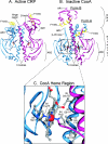
Comparison of the structures of active CRP and inactive CooA. (A) Active CRP (Protein Data Bank no. 1G6N) is a symmetrical homodimer that is rotated slightly here to display critical features. The two monomers are colored differently, and the upper portion of each constitutes the DNA-binding domain, although this is difficult to see in this protein species. The F helices, which make specific DNA contact, are depicted in yellow, and other important helices are indicated. The approximate positions of the AR1 and AR2 regions are indicated on onemonomer, and the AR3 region is circled on the other. cAMP is indicated by the pair of the ball-and-stick molecules near the center of each monomer. (B) The X-ray crystal structure of inactive CooA (Protein Data Bank no. 1FT9) shows that this protein exists in two forms, with the more extended form termed form B, although in each form the F helices are buried away from the solvent. DNA- and effector-binding domains are roughly indicated on the right side of the panel. Because of the extended structure, the AR3 region, at the tip of the β-4/5 loop, is easily seen. The heme region is depicted as the ball-and-stick structure and enlarged in panel C. (C) The heme vicinity of one CooA monomer is shown, with nearby residues noted and identified as to the protein monomer in which they are found: (a) refers to the monomer on the left, while (b) refers to the monomer on the right. His77 is shown as the ligand in the ferrous form, and Cys75; the ligand in the oxidized form, lies immediately behind it in this view. The other ligand, Pro2, is the N terminus of the right protein monomer and is connected to the red chain at the bottom right of this panel.
FIG. 4.
FIG. 4.
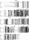
Sequence alignment of the CooA homologs (reprinted from reference 147). This shows the sequence comparison of the CooA homologs described in the text, with CooA of R. rubrum shown at the top and, for comparison, the CRP of E. coli at the bottom. Residues that are extremely or modestly conserved among the CooA homologs are shaded in black and gray, respectively. Above the top line, specific α-helix or β-sheet regions are noted, as are the following important regions: the distal heme pocket, which is formed both by the N-terminal residues and those around positions 112 to 117 in R. rubrum CooA (dotted lines); the proximal heme pocket, including Cys75 and His77 (dotted lines); the β-4 and β-5 regions forming the 4/5 loop and flanking the AR3 region; the portion of CooA deleted relative to CRP (termed “Gap”), which provides space for the heme; the hinge region with Phe132, which separates the effector- and DNA-binding domains; AR1, by analogy to a critical residue in CRP; and the E and F α helices, which define the DNA-binding region, with Gln178 shown near the beginning of the F helix. Rr, R. rubrum; Dh, D. hafniense; Ch, C. hydrogenoformans; Av, A. vinelandii; Dd, D. desulfuricans; Dv, D. vulgaris; Ec, E. coli.
FIG. 5.
FIG. 5.
Structure of the heme in the reduced form of CooA, oriented to show the relative positions of Cys75, which serves as the ligand in the oxidized form, and His77, which serves as the ligand in the reduced form. The Cys75 and heme Fe must move 2 to 3 Å with respect to each other for this ligation to occur.
FIG. 6.
FIG. 6.
The positioning of CooA with respect to RNA polymerase indicates that the two CooA monomers have fundamentally different interactions at class II promoters. The upstream monomer makes contact with the carboxyl-terminal domain of α (α-CTD), at the AR1 region, while the downstream monomer contacts the σ subunit at AR3 and the amino-terminal domain of α (α-NTD) at AR2.

References

    1. Abelsohn, A., M. D. Sanborn, B. J. Jessiman, and E. Weir. 2002. Identifying and managing adverse environmental health effects. 6. Carbon monoxide poisoning. CMAJ 166:1685-1690. - PMC - PubMed
    1. Adams, M. W. W., and E. I. Stiefel. 2000. Organometallic iron: the key to biological hydrogen metabolism. Curr. Opin. Chem. Biol. 4:214-220. - PubMed
    1. Akimoto, S., A. Tanaka, K. Nakamura, Y. Shiro, and H. Nakamura. 2003. O2-specific regulation of the ferrous heme-based sensor kinase FixL from Sinorhizobium meliloti and its aberrant inactivation in the ferric form. Biochem. Biophys. Res. Commun. 304:136-142. - PubMed
    1. Albracht, S. P. J., and R. Hedderich. 2000. Learning from hydrogenases: location of a proton pump and of a second FMN in bovine NADH-ubiquinone oxidoreductase (complex I). FEBS Lett. 485:1-6. - PubMed
    1. Aono, S. 2003. Biochemical and biophysical properties of the CO-sensing transcriptional activator CooA. Acc. Chem. Res. 36:825-831. - PubMed

Publication types

MeSH terms

LinkOut - more resources