. 2004 Sep 14;101(37):13636-41.
doi: 10.1073/pnas.0403998101. Epub 2004 Sep 7.

A nonpolio enterovirus with respiratory tropism causes poliomyelitis in intercellular adhesion molecule 1 transgenic mice

Affiliations

A nonpolio enterovirus with respiratory tropism causes poliomyelitis in intercellular adhesion molecule 1 transgenic mice

Andrew T Dufresne et al. Proc Natl Acad Sci U S A. .

Abstract

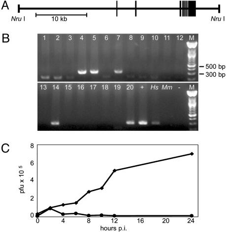
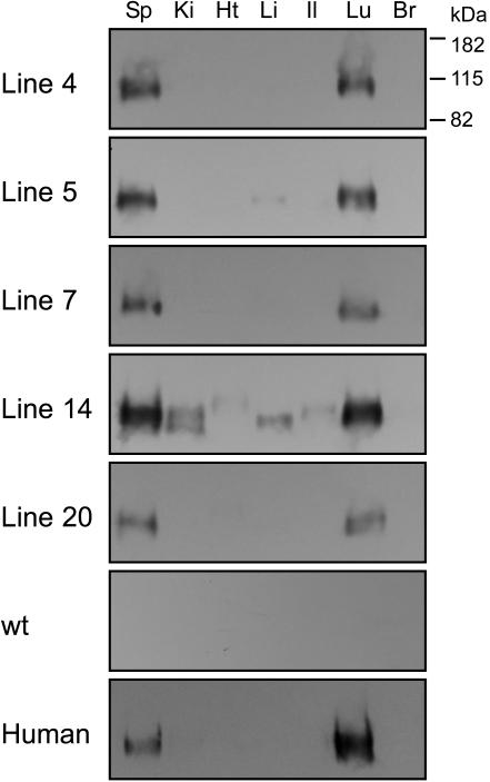
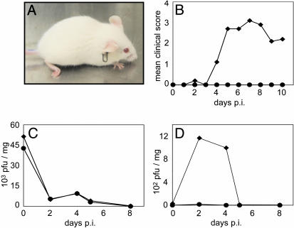
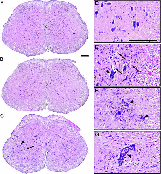
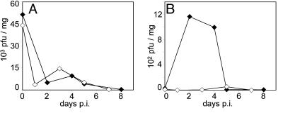
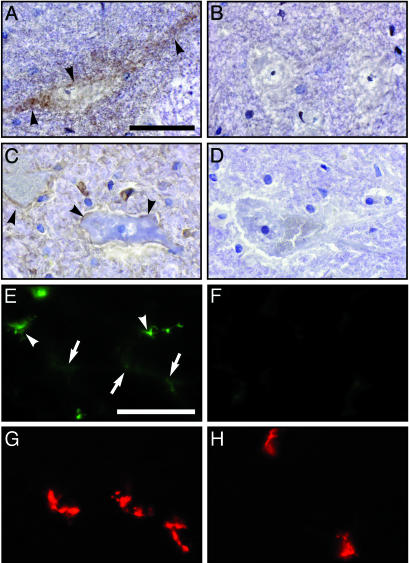
Coxsackievirus A21 (CAV21) is classified within the species Human enterovirus C (HEV-C) of the Enterovirus genus of picornaviruses. HEV-C share striking homology with the polioviruses (PV), their closest kin among the enteroviruses. Despite a high level of sequence identity, CAV21 and PV cause distinct clinical disease typically attributed to their differential use of host receptors. PV cause poliomyelitis, whereas CAV21 shares a receptor and a propensity to cause upper respiratory tract infections with the major group rhinoviruses. As a model for CAV21 infection, we have developed transgenic mice that express human intercellular adhesion molecule 1, the cell-surface receptor for CAV21. Surprisingly, CAV21 administered to these mice via the intramuscular route causes a paralytic condition consistent with poliomyelitis. The virus appears to invade the CNS by retrograde axonal transport, as has been demonstrated to occur in analogous PV infections. We detected human intercellular adhesion molecule 1 expression on both transgenic mouse and human spinal cord anterior horn motor neurons, indicating that members of HEV-C may share PV's potential to elicit poliomyelitis in humans.


Figures

Fig. 1.
Fig. 1.
Generation of tg mice. (A) Schematic 5′–3′ representation of the 39-kb DNA fragment containing the HsICAM-1 gene used for microinjection to derive tg mice (boxes signify HsICAM-1 exons). (B) PCR from founder genomic DNA (lanes 1–20) yields a specific 312-bp product for tg animals. +, BAC DNA; Hs, human genomic DNA; Mm, wild-type mouse genomic DNA; –, no template. (C) CAV21 replication in embryo fibroblast cultures established from HsICAM-1 tg embryos (♦) and non-tg littermates (•).
Fig. 2.
Fig. 2.
Antigen capture/immunoblot detection of HsICAM-1 in tissue homogenates from HsICAM-1 tg mouse lines 4, 5, 7, 14, and 20, a wild-type mouse, and corresponding human tissues. Sp, spleen; Ki, kidney; Ht, heart; Li, liver; Il, ileum; Lu, lung; Br, brain.
Fig. 3.
Fig. 3.
CAV21 pathogenicity in HsICAM-1 tg mice. (A) tg mouse 7 days after i.m. CAV21 injection. Note the characteristic drop-foot posture of the right hind limb. (B) Average clinical progression of CAV21-induced poliomyelitis in 10 tg mice (♦) and 8 wild-type controls (•). Severity of paresis was scored on an arbitrary scale (0, no symptoms; 1, minor lower paresis; 2, slight gait abnormality; 3, severe lower paresis/gait abnormality/drop foot; 4, ipsilateral paralysis). (C and D) CAV21 titers in gastrocnemius muscle (C) and spinal cord (D) of tg (♦) and wild-type mice (•) infected i.m. with CAV21 and killed at the indicated intervals.
Fig. 4.
Fig. 4.
Histopathology of CAV21-mediated poliomyelitis in the lumbar spinal cord of HsICAM-1 tg mice. (A and B) The spinal cords of mock-infected (A) and CAV21-infected non-tg (B) mice were histologically normal. (C) In the spinal cord of CAV21-infected HsICAM-1 tg mice, motor neuron destruction occurred ipsilaterally. Although contralateral motor neurons mostly remained unscathed (arrow), there was evidence of cellular infiltrates in their vicinity (arrowhead). (D–G) Histological detail of the anterior horn of CAV21-infected wild-type (D) and HsICAM-1 tg (E–G) mice. The latter had prominent evidence of neuronophagia (E and F, arrowheads) and eosinophilic bodies suggesting destroyed motor neurons (E, arrows). Mononuclear infiltrates occurred both diffusely in the anterior horn (C), surrounding affected neurons (E and F), and as perivascular cuffs (G, arrowhead). (Bars = 200 μm.)
Fig. 5.
Fig. 5.
CAV21 titers in gastrocnemius muscle (A) and spinal cord (B) of i.m.-infected tg mice with intact (♦) and severed (⋄) sciatic nerve.
Fig. 6.
Fig. 6.
Immunohistochemical detection of HsICAM-1. HsICAM-1 staining in the ventral horn of HsICAM-1 tg mouse (A) and human (C) lumbar spinal cord. Specificity was controlled by staining parallel sections from wild-type mice (B) and by incubating sections of human spinal cord with isotype-matched nonspecific primary antibody (D). HsICAM-1 signal was visualized with diaminobenzidine substrate (brown; arrowheads), and sections were counterstained with hematoxylin (blue). Fluorescent detection of HsICAM-1 (E and F) and NMJ (G and H) in gastrocnemius muscle of tg (E and G) and wild-type (F and H) mice. Two patterns of HsICAM-1 signal appeared in muscle (E): bright staining corresponding to capillary endothelium (arrowheads) and a more delicate signal that colocalized with NMJ (arrows; compare E and G). (Bars = 50 μm.)

References

    1. Grist, N. R., Bell, E. J. & Assaad, A. (1978) Prog. Med. Virol. 24, 114–157. - PubMed
    1. Dalldorf, G. & Melnick, J. L. (1965) in Viral and Rickettsial Infections of Man, eds. Horsfall, F. L & Tamm, I. (Lippincott, Philadelphia), 4th Ed., pp. 474–512.
    1. King, A. M. Q., Brown, F., Christian, P., Hovi, T., Hyypia, T., Knowles, N. J., Lemon, S. M., Minor, P. D., Palmenberg, A. C., Skern, T., et al. (2000) in Virus Taxonomy, Seventh Report of the International Committee on Taxonomy of Viruses, eds. Van Regenmortel, M. H. V., Fanquet, C. M., Bishop, D. H. L., Carstens, E. B., Estes, M. K., Lemon, S. M., Maniloff, J., Mayo, M. A., McGeoch, D. J., Pringle, C. R., et al. (Academic, San Diego), pp. 657–678.
    1. Pulli, T., Koskimies, P. & Hyypia, T. (1995) Virology 212, 30–38. - PubMed
    1. Pöyry, T., Kinnunen, L., Hyypiä, T., Brown, B., Horsnell, C., Hovi, T. & Stanway, G. (1996) J. Gen. Virol. 77, 1699–1717. - PubMed

Publication types

MeSH terms

Substances