. 2004 Sep 14;101(37):13436-41.
doi: 10.1073/pnas.0405868101. Epub 2004 Sep 7.

The crystal structure of ribosomal chaperone trigger factor from Vibrio cholerae

Affiliations

The crystal structure of ribosomal chaperone trigger factor from Vibrio cholerae

Anthony V Ludlam et al. Proc Natl Acad Sci U S A. .

Abstract

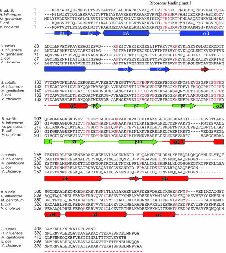
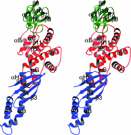
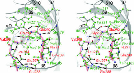
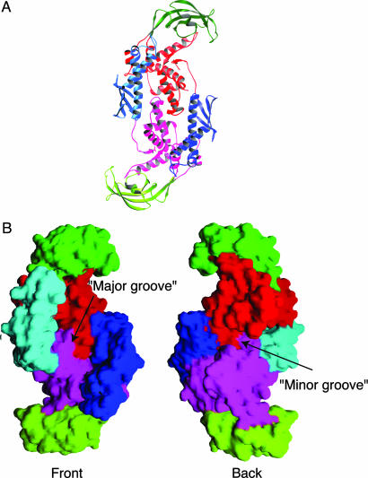
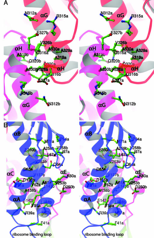
Trigger factor is a molecular chaperone that is present in all species of eubacteria. It binds to the ribosomal 50S subunit near the translation exit tunnel and is thought to be the first protein to interact with nascent polypeptides emerging from the ribosome. The chaperone has a peptidyl-prolyl cis-trans isomerase (PPIase) activity that catalyzes the rate-limiting proline isomerization in the protein-folding process. We have determined the crystal structure of nearly full-length trigger factor from Vibrio cholerae by x-ray crystallography at 2.5-A resolution. The structure is composed of two trigger-factor molecules related by a noncrystallographic two-fold symmetry axis. The monomer has an elongated shape and is folded into three domains: an N-terminal domain I that binds to the ribosome, a central domain II that contains PPIase activity, and a C-terminal domain III. The active site of the PPIase domain is occupied by a loop from domain III, suggesting that the PPIase activity of the protein could be regulated. The dimer interface is formed between domains I and III and contains residues of mixed properties. Further implications about dimerization, ribosome binding, and other functions of trigger factor are discussed.


Figures

Fig. 1.
Fig. 1.
Sequence alignment of bacterial trigger factors. The sequences of trigger factors from Bacillus subtilis, Haemophilus influenzae, Mycoplasma genitalium, E. coli, and V. cholerae were aligned by using clustalw (43). Invariant residues are indicated with red letters. Secondary structures are shown below the sequence: α-helices are drawn as cylinders, β-strands are drawn as arrows, other elements appear as solid lines, and structurally unobserved residues in the midsequence and at the C terminus are shown as dashed lines. Elements in domain I are blue, elements in domain II are green, and elements in domain III are red.
Fig. 2.
Fig. 2.
A stereo ribbon diagram of a trigger-factor monomer. Secondary structure elements in three domains of trigger factor are colored and labeled as in Fig. 1 (domain I, blue; domain II, green; and domain III, red). α-helices are drawn as coils, β-strands are drawn as arrows, and other elements are drawn as tubes. This figure was prepared with the program ribbons (44).
Fig. 3.
Fig. 3.
Interaction between the PPIase active site of domain II and the insertion loop from domain III. A close-up stereo view of the PPIase active site is shown. Domain II (PPIase) is shown as gray ribbon drawings. Side chains of residues that form the substrate-binding pocket are shown as ball-and-stick models (labeled in green). Note the overall hydrophobic nature of the pocket. The entire insertion loop (Glu-288 to Asn-298) is also shown in ball-and-stick rendition (labeled in red) superimposed with a light-red coil to facilitate chain tracing. Salt bridges and hydrogen bonds are depicted as dashed lines. Note that the catalytically important Tyr-221 makes a main chain contact with the amide group of Gly-293. This figure was prepared with the program ribbons (44).
Fig. 4.
Fig. 4.
The structure of the trigger-factor dimer. (A) A ribbon drawing of the trigger-factor dimer looking down the twofold axis of symmetry. Secondary structure elements in one of the trigger-factor subunits are colored as in Fig. 2. Rotating this subunit ≈90° around a vertical axis (bring the left half into the page and the right half out of the page) produces the molecule in Fig. 2. Corresponding elements in the other subunit are colored similarly but in a slightly different shade. This figure was prepared with the program ribbons (44). (B) Molecular surface representation of the trigger factor dimer. (Left) Front view of the molecule (same orientation as in A). (Right) Back view of the molecule. Two grooves on the surface of the molecule are indicated. This figure was prepared with the program grasp (45).
Fig. 5.
Fig. 5.
The dimer interface of trigger factor. (A) An enlarged stereo view of the residues involved in interactions between αG-αH helix–turn–helix motifs of two symmetry-related domains III. (B) An enlarged stereo view of residues involved in the dimer interface between helices αA and αB of domain I of one subunit, and helices αC and αE of domain III of the other subunit. Only residues that are involved in salt bridges, hydrogen bonds, or van der Waals interactions are shown. Note the position of the ribosome-binding loop relative to the dimer interface. Both figures were prepared with the program ribbons (44).

Similar articles

Cited by

References

    1. Hartl, F. U. & Hayer-Hartl, M. (2002) Science 295, 1852–1858. - PubMed
    1. Bukau, B., Deuerling, E., Pfund, C. & Craig, E. A. (2000) Cell 101, 119–122. - PubMed
    1. Lecker, S., Lill, R., Ziegelhoffer, T., Georgopoulos, C., Bassford, P. J., Jr., Kumamoto, C. A. & Wickner, W. (1989) EMBO J. 8, 2703–2709. - PMC - PubMed
    1. Hesterkamp, T., Hauser, S., Lutcke, H. & Bukau, B. (1996) Proc. Natl. Acad. Sci. USA 93, 4437–4441. - PMC - PubMed
    1. Huang, G. C., Li, Z. Y., Zhou, J. M. & Fischer, G. (2000) Protein Sci. 9, 1254–1261. - PMC - PubMed

Publication types

MeSH terms

Associated data

LinkOut - more resources