. 2004 Sep 3;305(5689):1466-70.
doi: 10.1126/science.1099191.

Activation of apoptosis in vivo by a hydrocarbon-stapled BH3 helix

Affiliations

Activation of apoptosis in vivo by a hydrocarbon-stapled BH3 helix

Loren D Walensky et al. Science. .

Abstract

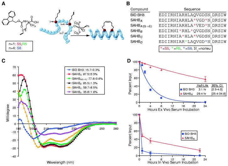
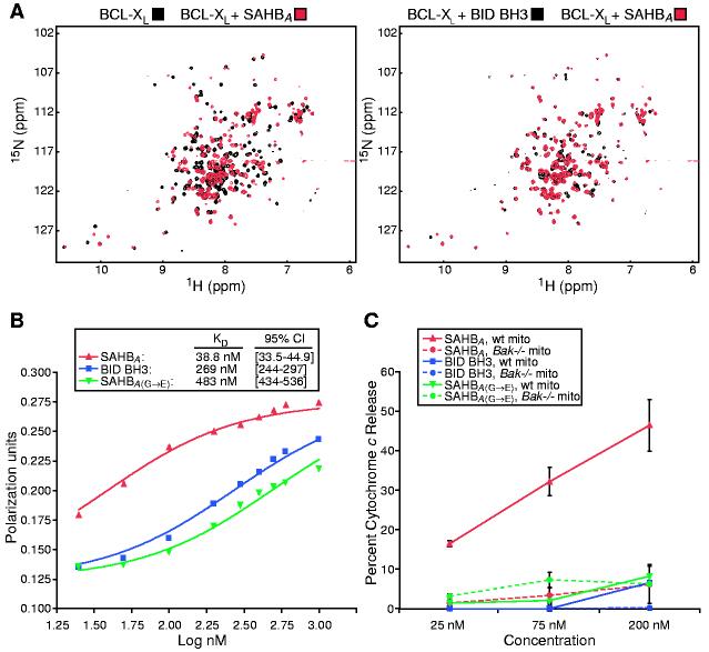
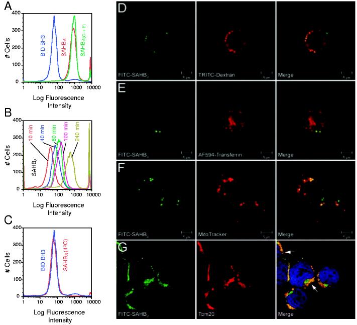
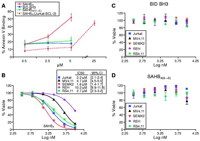
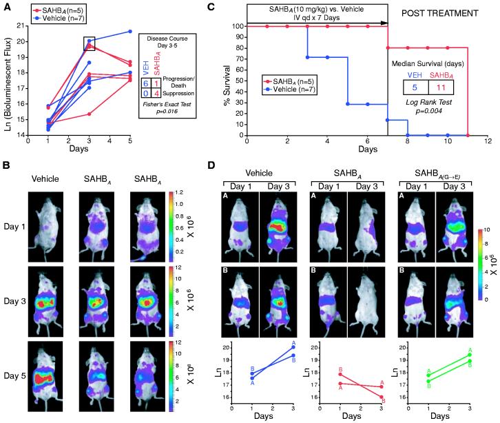
BCL-2 family proteins constitute a critical control point for the regulation of apoptosis. Protein interaction between BCL-2 members is a prominent mechanism of control and is mediated through the amphipathic alpha-helical BH3 segment, an essential death domain. We used a chemical strategy, termed hydrocarbon stapling, to generate BH3 peptides with improved pharmacologic properties. The stapled peptides, called "stabilized alpha-helix of BCL-2 domains" (SAHBs), proved to be helical, protease-resistant, and cell-permeable molecules that bound with increased affinity to multidomain BCL-2 member pockets. A SAHB of the BH3 domain from the BID protein specifically activated the apoptotic pathway to kill leukemia cells. In addition, SAHB effectively inhibited the growth of human leukemia xenografts in vivo. Hydrocarbon stapling of native peptides may provide a useful strategy for experimental and therapeutic modulation of protein-protein interactions in many signaling pathways.


Figures

Fig. 1.
Fig. 1.
Enhanced helicity, protease resistance, and serum stability of hydrocarbon-stapled BID BH3 compounds. (A and B) α,α-disubstituted non-natural amino acids containing olefinic side chains of varying length were synthesized as previously reported (, , ). Non-natural amino acid substitutions were made to flank three (substitution positions i and i+4) or six (i and i+7) amino acids within the BID BH3 peptide, so that reactive olefinic residues would reside on the same face of the α helix. (C) Circular dichroism was used to measure the percentages of SAHB maintained in helical configuration when dissolved in aqueous potassium phosphate solution (pH7) (supporting online material). (D) Fluoresceinated SAHBA and BID BH3 peptide were incubated at 37°C in mouse serum or injected intravenously (10 mg/kg) into NOD SCID mice. Serum concentrations of SAHBA and BID BH3 peptide were measured at the indicated time points with a fluorescence-based high-performance liquid chromatography detection assay. Both assays demonstrated enhanced serum stability of SAHBA.
Fig. 2.
Fig. 2.
SAHBA targets the binding pocket of BCL-XL, displays enhanced BCL-2 binding affinity, and specifically activates cytochrome c release from mitochondria in vitro. (A) HSQC experiments show similar spectral changes in 15N-BCL-XL upon binding SAHBA or BID BH3 peptide. (B) Kd's for binding of individual peptides to glutathione S-transferase-BCL-2 were determined by fluorescence polarization. (C) Mouse liver mitochondria (wild-type or Bak-/-, 0.5 mg/ml) were incubated for 40 min with 25 to 200 nM concentrations of BID BH3 peptide, SAHBA, or SAHBA(G→E), and cytochrome c was measured in the supernatant and sedimented mitochondria by an enzyme-linked immunosorbent assay.
Fig. 3.
Fig. 3.
SAHBA penetrates Jurkat leukemia cells by fluid-phase endocytosis and localizes to the mitochondrial membrane. Jurkat leukemia cells were incubated with FITC-labeled peptides for 4 hours at 37°C, followed by FACS analysis (A). FITC-SAHBA uptake occurred in a time-dependent manner at 37°C (B), but no FITC-SAHBA labeling was evident by 4 hours, when the experiment was performed at 4°C (C). Live confocal images demonstrated a colocalization of FITC-SAHBA with 4.4-kD dextran-labeled endosomes (D) but not transferrin-labeled endosomes (E) at 4 hours. A mitochondrial colocalization was evident by 24 hours, as demonstrated by the merged images of FITC-SAHBA and MitoTracker in live cells (F) and those of FITC-SAHBA and Tom20 (a mitochondrial outer-membrane marker) in fixed cells (G). Arrows highlight sites of colocalization corresponding to the surface of mitochondria cut in cross section (G).
Fig. 4.
Fig. 4.
SAHBA triggers apoptosis in Jurkat cells and inhibits a panel of human leukemia cells. FACS analysis of annexin V-treated cells was used to monitor apoptosis of Jurkat cells treated with 0.5 to 5 μM concentrations of BID BH3 peptide, SAHBA, or SAHBA(G→E) for 20 hours (A). Jurkat, REH, MV4;11, SEMK2, and RS4;11 leukemia cells were treated with serial dilutions of SAHBA (B), BID BH3 peptide (C), or SAHBA(G→E) (D), and MTT assays were performed at 48 hours to measure viability.
Fig. 5.
Fig. 5.
SAHBA suppresses growth of human leukemia cells in vivo, prolonging the survival of leukemic mice. (A) Leukemic SCID beige mice [with a day-1 natural logarithm (ln) bioluminescence range of 14.4 to 15.9] were treated with intravenous injections of 10 mg/kg SAHBA or vehicle (5% DMSO in D5W) daily for 7 days and were monitored for survival; leukemia burden was quantified by total body luminescence (photons/s/mouse) on days 1, 3, and 5. The disease course from days 3 to 5 differed between SAHBA-treated animals and controls (P = 0.016, Fisher's exact test [box in (A)], as illustrated by representative Xenogen images of bioluminescent leukemic mice (B); red signal represents the highest level of leukemia on the colorimetric scale. (C) Median survival was prolonged in SAHBA-treated animals as compared to controls (P = 0.004, log rank test). (D) To compare SAHBA with SAHBA(G→E), leukemic mice (with a day 1 ln bioluminescence range of 17.1 to 17.9) were treated daily with SAHB (10 mg/kg) or vehicle, and animals were imaged on days 1 and 3 to measure total body luminescence.

Comment in

Similar articles

Cited by

References

    1. Bakhshi A, et al. Cell. 1985;41:899. - PubMed
    1. Cleary ML, Sklar J. Proc. Natl. Acad. Sci. U.S.A. 1985;82:7439. - PMC - PubMed
    1. Tsujimoto Y, Cossman J, Jaffe E, Croce CM. Science. 1985;228:1440. - PubMed
    1. Danial NN, Korsmeyer SJ. Cell. 2004;116:205. - PubMed
    1. Wei MC, et al. Science. 2001;292:727. - PMC - PubMed

Publication types

MeSH terms