Latent effects of fibronectin, alpha5beta1 integrin, alphaVbeta5 integrin and the cytoskeleton regulate pancreatic carcinoma cell IL-8 secretion
- PMID: 15354211
- PMCID: PMC2409896
- DOI: 10.1038/sj.bjc.6602132
Latent effects of fibronectin, alpha5beta1 integrin, alphaVbeta5 integrin and the cytoskeleton regulate pancreatic carcinoma cell IL-8 secretion
Abstract
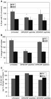
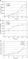
Interactions between tumour cells and the microenvironment are increasingly recognised to have an influence on cancer progression. In pancreatic carcinoma, a highly desmoplastic stroma with abnormal extracellular matrix (ECM) protein and interleukin-8 (IL-8) expression is seen. To investigate whether the ECM may further contribute to abnormalities in the microenvironment by influencing IL-8 secretion, we cultured the Mia PaCa2 pancreatic carcinoma cell line on fibronectin. This resulted in a dose-dependent increase in IL-8 secretion, which was RGD dependent and accompanied by cell spreading and proliferation. The role of spreading was assessed by disruption of the cytoskeleton with cytochalasin D, resulting in a large increase in IL-8 secretion, which was reduced from 31- to 24-fold by fibronectin. This remarkable response was associated with inhibition of spreading and proliferation and represents a novel cytoskeletal function. To investigate whether it could be accounted for by the loss of integrin-mediated signalling, the expressed alpha5beta1, alphaVbeta5 and alpha3beta1 integrins were inhibited. alpha5beta1 inhibition prevented spreading and proliferation but produced a much smaller rise in IL-8 secretion than cytochalasin D. alphaVbeta5 inhibition alone had only minor effects but when inhibited in combination with alpha5beta1 completely abolished the response to fibronectin. These results reveal latent stimulatory effects of the alphaVbeta5 integrin on IL-8 secretion and suggest that integrin crosstalk may limit the induction of IL-8 secretion by fibronectin. However, the magnitude of IL-8 secretion induced by cytochalasin cannot be accounted for by integrin signalling and may reflect the influence of another signalling pathway or a novel, intrinsic cytoskeletal function.
Figures


Similar articles
-
Fibronectin promotes brain capillary endothelial cell survival and proliferation through alpha5beta1 and alphavbeta3 integrins via MAP kinase signalling.J Neurochem. 2006 Jan;96(1):148-59. doi: 10.1111/j.1471-4159.2005.03521.x. Epub 2005 Nov 3. J Neurochem. 2006. PMID: 16269008
-
Role of alphavbeta5 integrins and vitronectin in Pseudomonas aeruginosa PAK interaction with A549 respiratory cells.Microbes Infect. 2004 Aug;6(10):875-81. doi: 10.1016/j.micinf.2004.05.004. Microbes Infect. 2004. PMID: 15310463
-
Role of α5β1 and αvβ3 integrins in relation to adhesion and spreading dynamics of prostate cancer cells interacting with fibronectin under in vitro conditions.Cell Biol Int. 2012 Oct 1;36(10):883-92. doi: 10.1042/CBI20110522. Cell Biol Int. 2012. PMID: 22686483
-
Integrins in cell adhesion and signaling.Hum Cell. 1996 Sep;9(3):181-6. Hum Cell. 1996. PMID: 9183647 Review.
-
Integrins: roles in cancer development and as treatment targets.Br J Cancer. 2004 Feb 9;90(3):561-5. doi: 10.1038/sj.bjc.6601576. Br J Cancer. 2004. PMID: 14760364 Free PMC article. Review.
Cited by
-
Integrin-mediated laminin-1 adhesion upregulates CXCR4 and IL-8 expression in pancreatic cancer cells.Surgery. 2007 Jun;141(6):804-14. doi: 10.1016/j.surg.2006.12.016. Epub 2007 May 4. Surgery. 2007. PMID: 17560257 Free PMC article.
-
Hydroxyapatite mineral enhances malignant potential in a tissue-engineered model of ductal carcinoma in situ (DCIS).Biomaterials. 2019 Dec;224:119489. doi: 10.1016/j.biomaterials.2019.119489. Epub 2019 Sep 11. Biomaterials. 2019. PMID: 31546097 Free PMC article.
-
Cancer cell angiogenic capability is regulated by 3D culture and integrin engagement.Proc Natl Acad Sci U S A. 2009 Jan 13;106(2):399-404. doi: 10.1073/pnas.0808932106. Epub 2009 Jan 6. Proc Natl Acad Sci U S A. 2009. PMID: 19126683 Free PMC article.
-
A 3D bioinspired highly porous polymeric scaffolding system for in vitro simulation of pancreatic ductal adenocarcinoma.RSC Adv. 2018 Jun 7;8(37):20928-20940. doi: 10.1039/c8ra02633e. eCollection 2018 Jun 5. RSC Adv. 2018. PMID: 35542351 Free PMC article.
-
Extracellular Influences: Molecular Subclasses and the Microenvironment in Pancreatic Cancer.Cancers (Basel). 2018 Jan 27;10(2):34. doi: 10.3390/cancers10020034. Cancers (Basel). 2018. PMID: 29382042 Free PMC article. Review.
References
-
- Anaya-Prado R, Toledo-Pereyra LH, Collins JT, Smejkal R, McClaren J, Crouch LD, Ward PA (1998) Dual blockade of P-selectin and beta2-integrin in the liver inflammatory response after uncontrolled hemorrhagic shock. J Am Coll Surg 187: 22–31 - PubMed
-
- Armstrong PB, Armstrong MT (2000) Intercellular invasion and the organizational stability of tissues: a role for fibronectin. Biochim Biophys Acta 1470: O9–20 - PubMed
-
- Bax DV, Bernard SE, Lomas A, Morgan A, Humphries J, Shuttleworth CA, Humphries MJ, Kielty CM (2003) Cell adhesion to fibrillin-1 molecules and microfibrils is mediated by alpha 5 beta 1 and alpha v beta 3 integrins. J Biol Chem 278: 34605–34616 - PubMed
MeSH terms
Substances
LinkOut - more resources
Full Text Sources
Medical

