. 2004 Nov;94(5):665-74.
doi: 10.1093/aob/mch191. Epub 2004 Sep 8.

The management of extracellular ice by petioles of frost-resistant herbaceous plants

Affiliations

The management of extracellular ice by petioles of frost-resistant herbaceous plants

M E McCully et al. Ann Bot. 2004 Nov.

Abstract

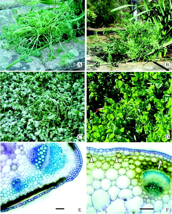
Background and aims: Some frost-tolerant herbaceous plants droop and wilt during frost events and recover turgor and posture on thawing. It has long been known that when plant tissues freeze, extracellular ice forms. Distributions of ice and water in frost-frozen and recovered petioles of Trifolium repens and Escholschzia californica were visualized.

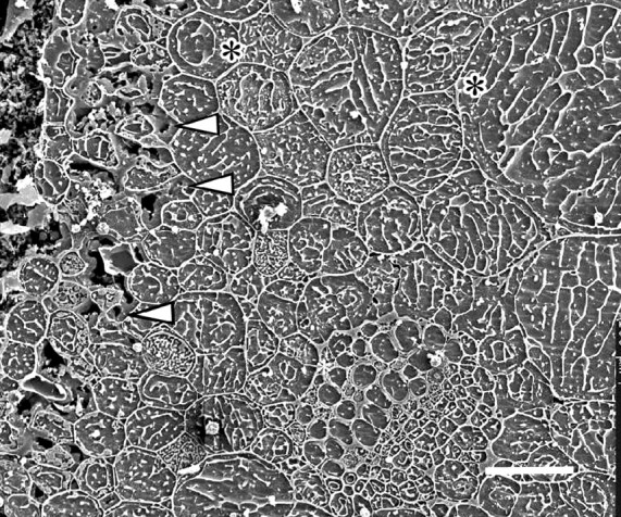
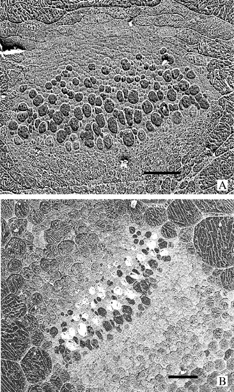
Methods: Petioles of intact plants were cryo-fixed, planed to smooth transverse faces, and examined in a cryo-SEM.

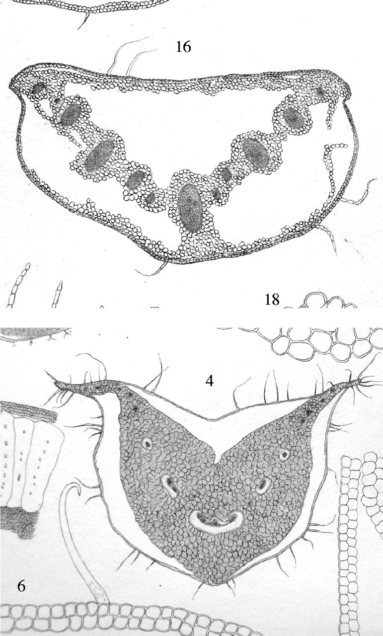
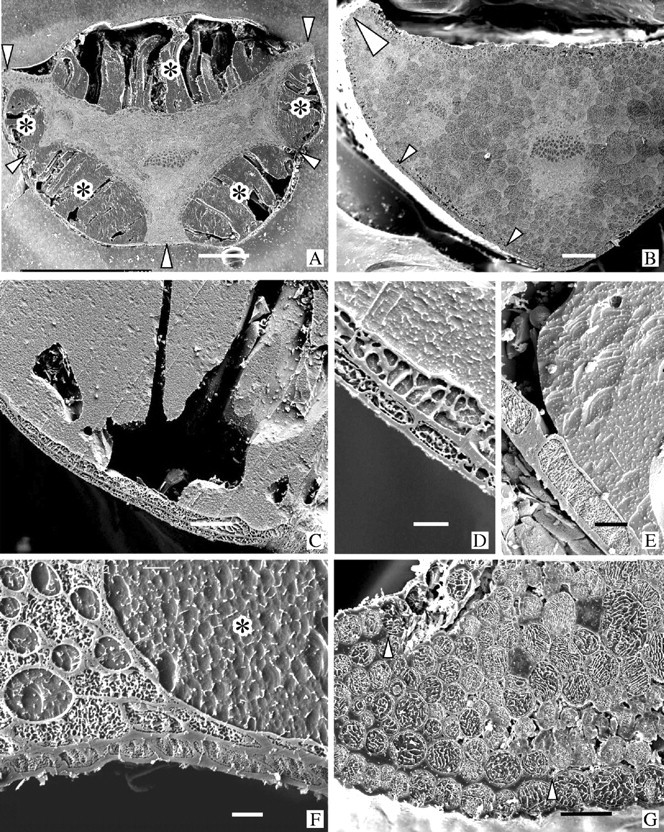
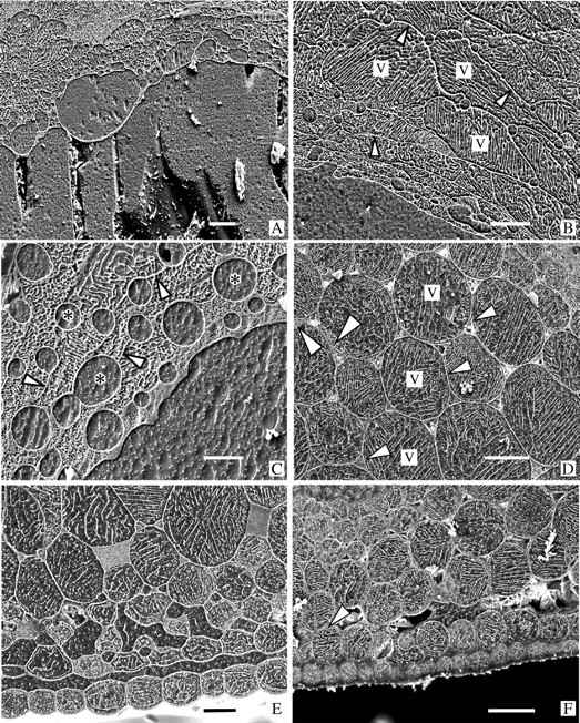
Key results: With frost-freezing, parenchyma tissues shrank to approx. one-third of their natural volume with marked cytorrhysis of the cells, and massive blocks of extracellular icicles grew under the epidermis (poppy) or epidermis and subepidermis (clover), leaving these layers intact but widely separated from the parenchyma except at specially structured anchorages overlying vascular bundles. On thawing, the extracellular ice was reabsorbed by the expanding parenchyma, and surface tissues again contacted the internal tissues at weak junctions (termed faults). These movements of water into and from the fault zones occurred repeatedly at each frost/thaw event, and are interpreted to explain the turgor changes that led to wilting and recovery. Ice accumulations at tri-cellular junctions with intercellular spaces distended these spaces into large cylinders, especially large in clover. Xylem vessels of frozen petioles were nearly all free of gas; in thawed petioles up to 20 % of vessels were gas-filled.

Conclusions: The occurrence of faults and anchorages may be expected to be widespread in frost-tolerant herbaceous plants, as a strategy accommodating extracellular ice deposits which prevent intracellular freezing and consequent membrane disruption, as well as preventing gross structural damage to the organs. The developmental processes that lead to this differentiation of separation of sheets of cells firmly cemented at determined regions at their edges, and their physiological consequences, will repay detailed investigation.


Figures

F<sc>ig</sc>. 1.
Fig. 1.
Photographs of two plates of drawings from Prillieux (1869) of transverse sections of frost-frozen petioles of Chelidonium quercifolium (upper drawing) and Symphytum tauricum (lower drawing). In both petioles large masses of ice had grown, drawing water from the shrunken parenchyma cells. The ice masses (seen here as spaces) remained within the distended intact outer tissue, which consisted of a few cell layers in C. quercifolium and a single epidermal layer in S. tauricum. Each petiole has three anchor points, where ice does not penetrate and the epidermis remains attached to the cortical tissues.
F<sc>ig</sc>. 2.
Fig. 2.
(A–D) Plants growing in late winter in Canberra. These and similar plants were used in this study. (A) California poppy at 07·00 h, temperature −4 °C. (B) The same plant at 08·30 h the same day when warmed by the sun, ambient temperature +4 °C. (C) White clover at 07·00 h, temperature −4°C. (D) Same patch of clover on the same day at 08·00 h after brief warming by the sun, ambient temperature +3 °C. (E and F) Hand-cut transverse sections of petioles stained with toluidine blue showing the inherent regions of weakness (faults) between surface tissues and the underlying chlorenchyma at the petiole periphery, and anchorage regions where surface tissues do not detach during extracellular ice formation. (E) California poppy. This petiole was sectioned in early summer and was from a young leaf that had never been frozen. During sectioning the single-layered lower epidermis separated at the fault over the chlorenchyma but not at the anchorage over the vascular bundle at the wing of the petiole. Bar = 100 μm. (F) White clover. This petiole was sectioned in August and was from a leaf that had experienced at least six freezing/thawing events previously. Arrowheads indicate the fault separating the two surface layers from the underlying chlorenchyma. The arrow indicates the anchorage over the vascular bundle. Bar = 100 μm.
F<sc>ig</sc>. 3.
Fig. 3.
Transverse faces of cryo-planed sections of cryo-fixed petioles viewed in a CSEM. Specimens in A, C–E and F were frost-frozen; B and G were thawed. All were from the plants shown in Fig. 2A–D. In each micrograph the lower epidermis is at the bottom. (A) Poppy. Leaf was frost-frozen as in Fig. 2A. Water has been drawn out of the ground parenchyma which is now much shrunk and distorted, and has accumulated in large deposits of ice (dark grey, typical ones marked by *) on both the upper and lower sides of the petiole where the epidermis has separated from the underlying chlorenchyma. These ice deposits, though incredibly large, are always encompassed on their outer edge by the intact, separated epidermis. During cryo-planing portions of the ice deposits crack, drop out and are lost. Tight anchorages (arrowheads) between epidermis and underlying tissue over the vascular bundles prevent loss of the epidermis. Bar = 500 μm. (B) Poppy. Petiole from the same plant as in A but after recovery as shown in Fig. 2B. The ice has all melted and the extracellular water has been retrieved by the ground parenchyma cells that have recovered their turgor. The epidermis is in place against the underlying cells (small arrowheads). The large arrowhead indicates an anchorage at the petiole wing similar to that in G. Bar = 200 μm. (C) Clover. Surface tissues around the outside of a large extracellular ice deposit from a leaf shown in Fig. 2C. Bar = 50 μm. (D) Clover. Detail of a region similar to C showing the double-layered coherent surface tissue typical of clover. Bar = 10 μm. (E) Poppy. Region similar to that of C and D showing the single epidermal layer characteristic of the separating surface tissue in poppy. Bar = 20 μm. (F) Clover. An anchorage overlying thick-walled cells linking a vascular bundle to the epidermis in a frost-frozen petiole. Note the enlarged and rounded ice-filled intercellular spaces at the tricellular junctions. The asterisk marks a portion of a large peripheral ice deposit as in C. Bar = 10 μm. (G) Poppy. A thawed petiole showing the thick anchorage (arrowheads) at the tip of the wing (large arrowhead in B), and the loose attachment of the epidermis to the underlying cells at the fault (clearest under the three epidermal cells at the lower right). Bar = 50 μm.
F<sc>ig</sc>. 4.
Fig. 4.
All are cryo-preparations of petioles as in Fig. 3. The lower epidermis is at or beyond the bottom of each micrograph. A, B and C were frost-frozen; D, E and F were thawed. (A) Poppy. The upper surface of a large peripheral ice deposit as in Fig. 3A showing the intersection with the dehydrated and distorted parenchyma cells, and intercellular spaces that have been ballooned out by the accumulating ice during frost-freezing. Bar = 50 μm. (B) Poppy. Parenchyma cells bordering a large ice deposit showing typical variations in size and shape of the cells dehydrated during frost-freezing. Note the clearly defined, ice-filled intercellular spaces. Arrowheads indicate cell walls. v = vacuole. Bar = 50 μm. (C) Clover. A location similar to that in A. In this species the cells tend to be so shrunken by the frost-freezing that some walls are difficult to distinguish (arrowheads). The ice-filled intercellular spaces (*) are usually much larger than those in poppy. Bar = 20 μm. (D) Clover. Fully rehydrated, turgid parenchyma in a recovered petiole. The small triangular intercellular spaces are mostly gas filled (the white material in most of them is ice chips that fell into the spaces during planing). Tonoplasts (small arrowheads) and nuclei (large arrowheads) can be distinguished in some of the cells. v = vacuole. Bar = 50 μm. (E) Poppy. Reabsorption of extracellular water was not quite complete when this thawing petiole was cryo-fixed. The epidermis is still separated from the chlorenchyma by water and, although the inner parenchyma have partly returned to their turgid form, those near the surface are still shrunken and distorted, and intercellular spaces are water-filled. Bar = 30 μm. (F) Clover. Cells in this petiole are back to normal turgidity but a small amount of water is still free in the fault. Arrowhead indicates the beginning of an anchorage which extends beyond the left edge of the micrograph. Bar = 50 μm.
F<sc>ig</sc>. 5.
Fig. 5.
Clover. Cryo-preparation. A partially thawed region of a petiole where the inner parenchyma cells are returning to full turgor but still angular in outline and their intercellular spaces still water-filled. The outermost chlorenchyma cells (left of micrograph) are still very shrunken and distorted. Arrowheads indicate residual water in the fault region. The xylem vessels (lower right of centre) are all water-filled. A wide, watery space between the vacuole and cell wall (*) is often seen in cells at this stage of rehydration. Bar = 50 μm.
F<sc>ig</sc>. 6.
Fig. 6.
Cryo-preparations. (A) A large vascular bundle in a frost-frozen petiole of a poppy preparation as the one in Fig. 3A. The bundle is surrounded by collapsed parenchyma cells and all the xylem vessels are ice-filled. Bar = 50 μm. (B) A similar vascular bundle in a thawed petiole as seen in Fig. 3B. The surrounding parenchyma cells are rehydrated and many of the xylem vessels are embolized (they appear white because of the ice chips that lodge in the spaces during planing). Bar = 50 μm.

References

    1. Ashworth EN. 1990. The formation and distribution of ice within flower buds. Plant Physiology 92: 718–725. - PMC - PubMed
    1. Ashworth EN, Echlin P, Pearce RS, Hayes TL. 1988. Ice formation and tissue response in apple twigs. Plant, Cell and Environment 11: 703–710.
    1. Ashworth EN, Davis GA, Wisniewski ME. 1989. The formation and distribution of ice within dormant and deacclimated peach flower buds. Plant, Cell and Environment 12: 521–528.
    1. Ball MC, Canny MJ, Huang CX, Heady R. 2004. Structural changes in acclimated and unacclimated Eucalyptus leaves during freezing and thawing. Functional Plant Biology 31: 29–40. - PubMed
    1. Ball MC, Wolfe J, Canny M, Hofmann M, Nicotra AB, Hughes D. 2002. Space and time dependence of temperature and freezing in evergreen leaves. Functional Plant Biology 29: 1259–1272. - PubMed