. 2004 Sep 8;24(36):7804-13.
doi: 10.1523/JNEUROSCI.1941-04.2004.

Ipsilateral actions of feline corticospinal tract neurons on limb motoneurons

Affiliations

Ipsilateral actions of feline corticospinal tract neurons on limb motoneurons

S A Edgley et al. J Neurosci. .

Abstract

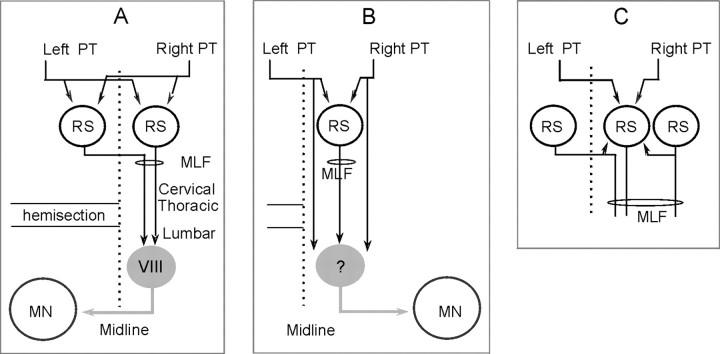
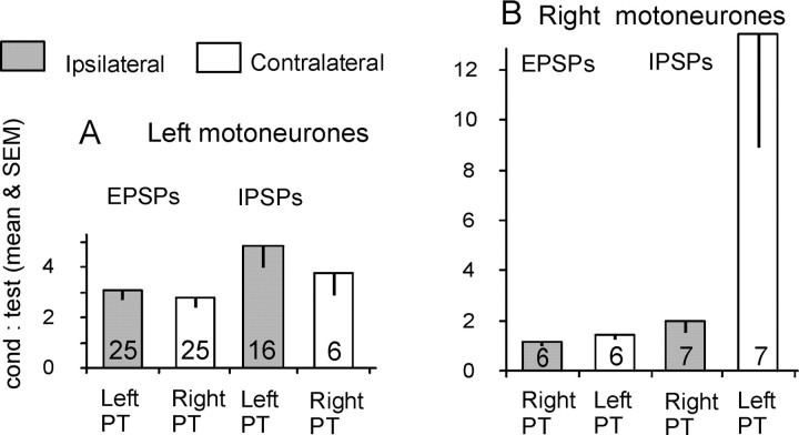
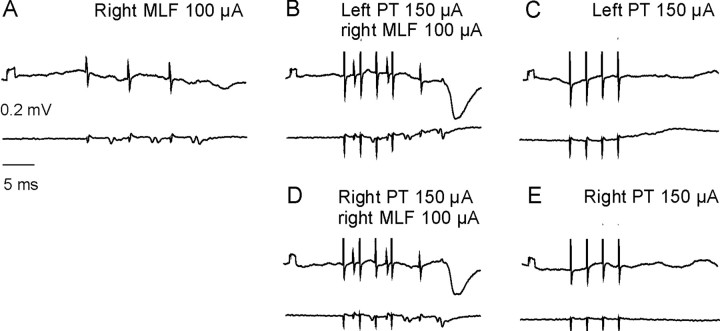
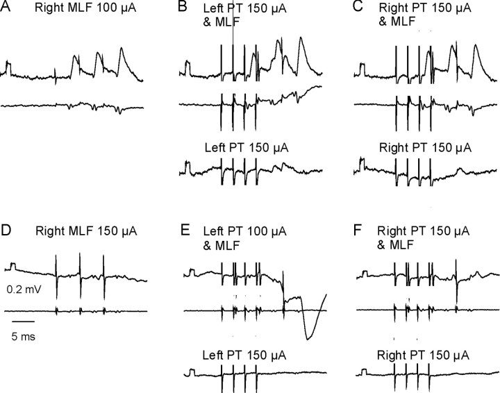
Contralateral pyramidal tract (PT) neurons arising in the primary motor cortex are the major route through which volitional limb movements are controlled. However, the contralateral hemiparesis that follows PT neuron injury on one side may be counteracted by ipsilateral of actions of PT neurons from the undamaged side. To investigate the spinal relays through which PT neurons may influence ipsilateral motoneurons, we analyzed the synaptic actions evoked by stimulation of the ipsilateral pyramid on hindlimb motoneurons after transecting the descending fibers of the contralateral PT at a low thoracic level. The results show that ipsilateral PT neurons can affect limb motoneurons trisynaptically by activating contralaterally descending reticulospinal neurons, which in turn activate spinal commissural interneurons that project back across to motoneurons ipsilateral to the stimulated pyramidal tract. Stimulation of the pyramids alone did not evoke synaptic actions in motoneurons but potently facilitated disynaptic EPSPs and IPSPs evoked by stimulation of reticulospinal tract fibers in the medial longitudinal fascicle. In parallel with this double-crossed pathway, corticospinal neurons could also evoke ipsilateral actions via ipsilateral descending reticulospinal tract fibers, acting through ipsilaterally located spinal interneurons. Because the actions mediated by commissural interneurons were found to be stronger than those of ipsilateral premotor interneurons, the study leads to the conclusion that ipsilateral actions of corticospinal neurons via commissural interneurons may provide a better opportunity for recovery of function in hemiparesis produced by corticospinal tract injury.


Figures

Figure 1.
Figure 1.
Diagrams showing hypothetical connections between PT fibers and ipsilateral hindlimb motoneurons (MN). A, Connections between left and right pyramidal tract fibers and motoneurons on the left side via left and right RS neurons with axons in the MLF and lumbar commissural interneurons. In preparations in which the left half of the spinal cord was transected at a low thoracic level, ipsilateral actions of left PT fibers on motoneurons could be mediated by RS neurons with axons descending in the right MLF. The direct corticospinal projection is omitted for the sake of simplicity. B is the same as A but for connections with motoneurons on the right side. Ipsilateral actions of right PT fibers on motoneurons are mediated by RS neurons with axons descending in the right MLF and local ipsilateral premotor interneurons indicated by a question mark. Intact corticospinal tract fibers on the right side of the spinal cord are added, but RS neurons located on the left side are omitted for the sake of simplicity. C, A diagram explaining facilitation of synaptic actions evoked from MLF actions by preceding PT stimuli, considering that PT fibers coexcite RS neurons with input from other neurons in the reticular formation and facilitate synaptic actions of these neurons. The diagram shows convergence on an RS neuron with an ipsilateral axon but would be equally valid for RS neurons with crossed descending projections and input from PT fibers of both sides.
Figure 2.
Figure 2.
Reconstructions of the locations of the stimulating electrodes. A-C show locations of electrode tips as defined by the electrolytic lesions made at the end of the experiments in the MLF and in the left and right PT, respectively. These are superimposed on representative sections of the brainstem cut in the plane of the electrode insertions. All MLF electrode placements were within the borders of the MLF, and all PT placements were within the pyramids or at the border with the TB. SO, Superior olive.
Figure 3.
Figure 3.
Descending volleys evoked by interacting stimulation of the right MLF and the left PT. A-C, Descending volleys recorded from the cord dorsum in the C4 segment (averages of 10 records). A shows the later components of volleys evoked by a train of PT stimuli (in the boxes) and the increase in the amplitudes of these components after each successive stimulus. B shows a similar increase in amplitude of the later components of descending volleys evoked by a train of MLF stimuli (in box). C illustrates a large increase in the amplitude of the late component of the volley evoked by the first MLF stimulus (in box), when the MLF stimuli were preceded by a train of PT stimuli. D and E show a similar facilitation of late components of descending MLF volleys recorded at thoracic level. In this and the following figures, the largest stimulus artifacts have been truncated.
Figure 4.
Figure 4.
Facilitation of the late components of MLF descending volleys and of disynaptic EPSPs in motoneurons by stimulation of both left and right PT after elimination of corticospinal tract fibers at C2-C3. A, Descending volleys after stimulation of the right MLF with a train of three stimuli, recorded at the Th12 level (averages of 10 records). The arrow indicates the abortive late component evoked by the first stimulus. B, Effects of conditioning stimuli in the left pyramids on the volleys evoked by a single MLF stimulus (equivalent to the first stimulus shown in A) showing a large late component (arrow). C and D are the same as B but after successively made lesions of the right and left DLF in the C2 segment, transecting connections between the left and right PT fibers and C3-C4 propriospinal neurons as indicated in the diagram. The extent of the lesions is shown in the two sections (∼4 mm apart rostrocaudally) to the left. The disappearance of the PT volleys is illustrated in E and F where gray and black traces are records obtained before and after the lesions together with the differences between them. E-H, Intracellular records from a GS motoneuron after lesions of the dorsolateral funiculus in the C2 segment (shown to the left of I and J, with the corresponding control records in K and L) in another experiment.
Figure 5.
Figure 5.
Effects of a stimulation of the left PT on descending volleys evoked by subsequent stimulation of the MLF. A and B plot the time course of changes in peak-to-peak amplitudes of volleys evoked by a single stimulus (or the first of a train) applied in the right MLF as a function of time intervals between these stimuli and the final stimulus of a train of conditioning PT stimuli (4 stimuli; 400 Hz) from two experiments. The x-axis labels refer to the time between the final PT stimulus and the MLF stimulus. The amplitudes are expressed as a ratio of the amplitudes of conditioned (cond) and nonconditioned (test) volleys and were measured from averages of 20 individual records. The black symbols represent the early components (E-H, boxes) and show changes attributable to the effects of the fourth PT stimulus. The open symbols show changes in the early components that are attributable to the third stimulus of the train to the PT. The gray symbols represent the late components. C and D show similar data for the largest changes in volleys recorded at the L6 and C4 segmental levels (from the same experiment as the plots in A and C, respectively). Note that in C, the time intervals for facilitation and occlusion evoked by the third and fourth stimulus are superimposed. Therefore, 0 on the abscissa corresponds in this plot to the time of application of the third stimulus (open symbols) and the fourth PT stimulus (filled symbols). E, F, Examples of test and conditioned volleys recorded at thoracic level at a conditioning testing interval, indicated by the double arrow, at which the early components of the volleys following the first MLF stimulus (boxed) were most effectively depressed. G, H, Test and conditioned volleys simultaneously recorded at cervical level. Voll., Volley.
Figure 6.
Figure 6.
Facilitation of disynaptic EPSPs evoked from the MLF by conditioning stimulation of the pyramids. Top records in each panel are intracellular potentials from a GS motoneuron on the left side of the spinal cord; the bottom records are from cord dorsum. Voltage calibration (0.2 mV for the calibration pulse at the beginning of the records) in B is for the intracellular records. The largest stimulus artifacts have been truncated. A, Test EPSPs evoked by stimulation of the right MLF with a train of three stimuli that were too weak to produce a temporal facilitation. B, Temporal facilitation of EPSPs evoked by stronger MLF stimuli. C, D, Facilitation of effects of weak MLF stimuli when the left (C) or right (D) PT were also stimulated with a train that terminated ∼5 msec before the final MLF stimulus. E, F, Negligible actions of stimuli to the left and right PT when stimulated alone.
Figure 7.
Figure 7.
Parameters of PT stimulation needed for effective facilitation of synaptic actions evoked from MLF. The top records in each panel are from a GS motoneuron on the left side of the spinal cord. The bottom records are from the cord dorsum. A-C, Small EPSPs were evoked in this motoneuron by a train of three MLF stimuli at 25 μA, and considerable temporal facilitation was evident with stronger stimulation (50 and 200 μA). D-F, Spatial facilitation of the EPSPs evoked by trains of stimuli to the PT at different frequencies. Note that three PT stimuli at 200 Hz evoked only marginal facilitation of the EPSP evoked by the third MLF stimulus, and that the effectiveness of the PT stimuli was much greater when their frequency increased from 200 to 300 Hz (E), and in particular when a fourth stimulus was added (F). Otherwise, the format is the same as in Figure 6.
Figure 8.
Figure 8.
Time course of facilitation of disynaptic PSPs evoked from MLF by stimulation of pyramidal tract fibers. A, B, Time course of facilitation of EPSPs evoked in GS motoneurons on the left side of the spinal cord after stimulation of the left and right pyramids indicated by gray and black symbols, respectively. C, Similar data for IPSPs evoked in another GS motoneuron. Intervals were measured between the fourth PT stimulus and the third MLF stimulus. The area of the PSPs was measured within 2 msec time windows from their onset. Time 0 on the x-axis represents the timing of the effective MLF stimulus, relative to the last PT conditioning stimulus. cond, Conditioned.
Figure 9.
Figure 9.
Comparison of the effectiveness of facilitation of PSPs evoked from the right MLF in motoneurons on the left (A) and right (B) side of the spinal cord by conditioning stimulation of the left and right PT. The bars represent the mean amount of facilitation [ratio of conditioned (cond) to test] with SE bars. A, Facilitation in motoneurons on the left (via commissural interneurons). B, Facilitation in motoneurons on the right (via ipsilaterally projecting neurons). Facilitatory actions of left and right pyramids (abscissa) were tested on the same motoneurons and using the same parameters of stimulation. The only statistically significant differences (Student's t test; p = 0.04) between the effects evoked from the left and right PT were those on IPSPs evoked in the right side motoneurons.
Figure 10.
Figure 10.
Facilitation of disynaptic IPSPs evoked from MLF by conditioning stimulation of pyramids. Top records in each panel are from a GS motoneuron on the left side. Bottom records are from cord dorsum. Otherwise, the format is the same as in Figure 6.
Figure 11.
Figure 11.
Facilitation of disynaptic EPSPs and IPSPs in motoneurons on the right side of the spinal cord. Top records in each panel are intracellular records from a motoneuron on the right side of the cord. A-C, From a GS motoneuron; D-F, from a Q motoneuron. Bottom records are from the cord dorsum. A and D show the responses of the motoneurons to a train of three stimuli to the MLF. B, C, E, and F show the effects of a train of PT stimuli delivered to end 5-10 msec before the final MLF stimulus. The lowermost records in B, C, E, and F are as the top records but showing effects of PT stimuli alone. Otherwise, the format is the same as in Figure 6 (the figure illustrates effects summarized in Figure 9B).

Similar articles

Cited by

References

    1. Alstermark B, Pinter M, Sasaki S (1983) Brainstem relay of disynaptic pyramidal EPSPs to neck motoneurons in the cat. Brain Res 259: 147-150. - PubMed
    1. Alstermark B, Lundberg A, Pinter M, Sasaki S (1987) Subpopulations and functions of long C3-C5 propriospinal neurones. Brain Res 404: 395-400. - PubMed
    1. Alstermark B, Pinter MJ, Sasaki S (1992) Descending pathways mediating disynaptic excitation of dorsal neck motoneurones in the cat: brain stem relay. Neurosci Res 15: 42-57. - PubMed
    1. Armand J, Kuypers HG (1980) Cells of origin of crossed and uncrossed corticospinal fibers in the cat: a quantitative horseradish peroxidase study. Exp Brain Res 40: 23-34. - PubMed
    1. Armand J, Holstege G, Kuypers HG (1985) Differential corticospinal projections in the cat. An autoradiographic tracing study. Brain Res 343: 351-355. - PubMed

Publication types

LinkOut - more resources