

doi: 10.1523/JNEUROSCI.2420-04.2004.
L1.1 is involved in spinal cord regeneration in adult zebrafish
Affiliations
- PMID: 15356195
- PMCID: PMC6729920
- DOI: 10.1523/JNEUROSCI.2420-04.2004
Item in Clipboard
L1.1 is involved in spinal cord regeneration in adult zebrafish
J Neurosci.
.
Abstract
Adult zebrafish, in contrast to mammals, regrow axons descending from the brainstem after spinal cord transection. L1.1, a homolog of the mammalian recognition molecule L1, is upregulated by brainstem neurons during axon regrowth. However, its functional relevance for regeneration is unclear. Here, we show with a novel morpholino-based approach that reducing L1.1 protein expression leads to impaired locomotor recovery as well as reduced regrowth and synapse formation of axons of supraspinal origin after spinal cord transection. This indicates that L1.1 contributes to successful regrowth of axons from the brainstem and locomotor recovery after spinal cord transection in adult zebrafish.
Figures
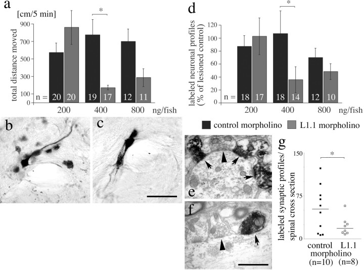
Morpholinos to L1.1 specifically impair locomotor and anatomical recovery 6 weeks after spinal cord transection. a, The total distance moved is reduced at 400 ng of L1.1 morpholino per fish, compared with control morpholino (*p < 0.05). b, c, Neuronal profiles in cross sections of the magnocellular octaval nucleus in the brainstem are back-labeled by HRP from a position caudal to the spinal transection site in control morpholino-treated (b) and to a lesser extent in L1.1 morpholino-treated (c) animals. Different focal planes are combined in the photomicrographs. d, The number of cell profiles back-labeled in the brainstem from a position caudal to a spinal transection site is significantly reduced after application of L1.1 morpholino compared with control morpholino at 400 ng per fish. Values are expressed as percentage of the labeled profiles counted in lesioned animals that had not received morpholino (*p < 0.05). e-g, The number of anterogradely traced synapses formed by regenerating axons from the brainstem caudal to a spinal transection site is reduced by L1.1 morpholino treatment. Labeled synapses of supraspinal origin (arrows) appear much darker than unlabeled synapses (arrowheads) of intraspinal neurons in the caudal spinal cord in control morpholino- (e) and L1.1 morpholino-treated (f) animals. In g, a graphic comparison of the numbers of labeled synapses found in individual animals is given (bars represent median values; *p < 0.05). Scale bars: (in c) b, c, 75 μm; (in f) e, f, 1 μm. Error bars represent SEM.
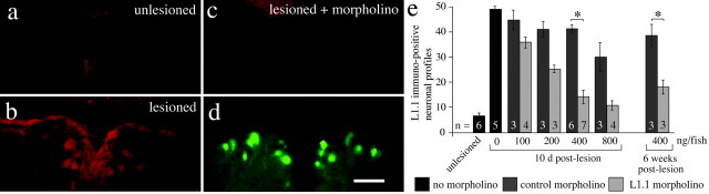
Morpholinos are retrogradely transported into neuronal somata in the brainstem and reduce L1.1 immunoreactivity. a-d, In a cross section through the NMLF, the number of L1.1-immunopositive neurons is increased 10 d after a lesion (b) compared with unlesioned controls (a). Upregulation is greatly reduced when L1.1 morpholino is applied to the spinal lesion site (c). In the same tissue section (d), accumulation of fluorescein-tagged L1.1 morpholino is visible with the appropriate filters. e, The number of L1.1-immunoreactive cell profiles is reduced by L1.1 morpholino compared with control morpholino-treated animals in a concentration-dependent manner (*p < 0.05). Scale bar: (in d) a-d, 50 μm. Error bars represent SEM.
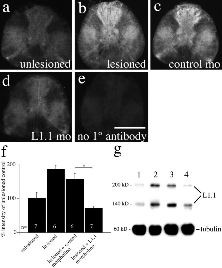
L1.1 morpholino reduces L1.1 immunolabeling caudal to a spinal transection site at 10 d postlesion. a-e, Immunofluorescence for L1.1 is increased 1 mm caudal to a spinal lesion site in lesioned animals (b) and lesioned animals that received control morpholino (c), but not in L1.1 morpholino (mo)-treated animals (d), compared with unlesioned controls (a) at 10 d postlesion. In e, the primary antibody (1°) was omitted on a spinal cross section from a lesioned animal. f, Quantification of L1.1 immunofluorescence in spinal cross sections 1 mm caudal to the lesion site indicates a significant reduction by L1.1 morpholino treatment compared with control morpholino treatment (*p < 0.05). g, Western blot analysis of the same spinal region indicates increased L1.1 immunoreactivity in a band at 200 kDa and another band at 140 kDa, which probably represents a proteolytic degradation product of L1.1, in lesioned animals (lane 2) and those that had received the control morpholino (lane 3) compared with unlesioned controls (lane 1). Upregulation of L1.1 was weaker in L1.1 morpholino-treated animals (lane 4). Anti-tubulin labeling served as a loading control. Scale bar: (in e) a-e, 100 μm. Error bars represent SEM.
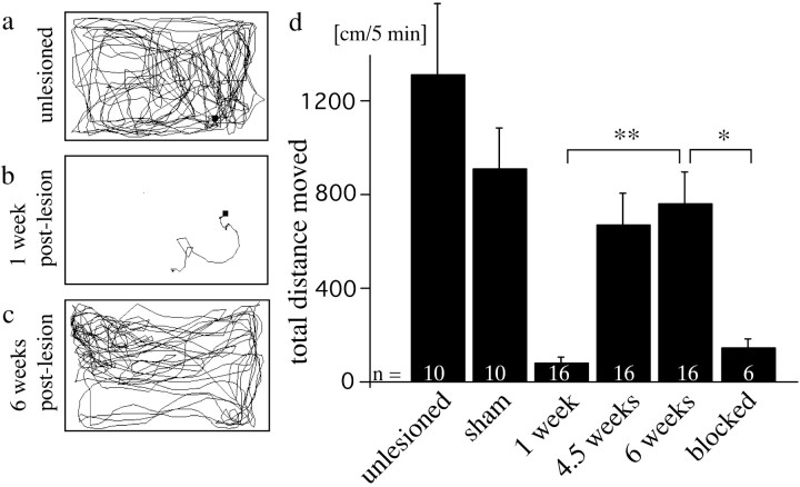
The total distance moved partially recovers after spinal cord transection. a, Swim track of an unlesioned fish. b, c, Swim tracks of the same fish, 1 week (b) and 6 weeks (c) after spinal cord transection, show recovery of total distance moved. d, The total distance moved at 6 weeks postlesion is significantly different from values at 1 week postlesion and from values for fish in which regeneration was mechanically blocked at 6 weeks postlesion (*p < 0.05; **p < 0.01). Values for fish in which only the muscle tissue, but not the spinal cord, was cut (sham) is indicated at 10 weeks postlesion. Error bars represent SEM.
References
-
- Asher RA, Morgenstern DA, Moon LD, Fawcett JW (2001) Chondroitin sulphate proteoglycans: inhibitory components of the glial scar. Prog Brain Res 132: 611-619. - PubMed
-
- Bareyre FM, Kerschensteiner M, Raineteau O, Mettenleiter TC, Weinmann O, Schwab ME (2004) The injured spinal cord spontaneously forms a new intraspinal circuit in adult rats. Nat Neurosci 7: 269-277. - PubMed
-
- Becker CG, Schweitzer J, Feldner J, Schachner M, Becker T (2004) Tenascin-R as a repellent guidance molecule for newly growing and regenerating optic axons in adult zebrafish. Mol Cell Neurosci 26: 376-389. - PubMed
-
- Becker T, Becker CG (2001) Regenerating descending axons preferentially reroute to the gray matter in the presence of a general macrophage/microglial reaction caudal to a spinal transection in adult zebrafish. J Comp Neurol 433: 131-147. - PubMed
Publication types
MeSH terms
Substances
LinkOut - more resources
Full Text Sources
Medical
Molecular Biology Databases