
Hereditary and acquired angioedema: problems and progress: proceedings of the third C1 esterase inhibitor deficiency workshop and beyond

Angelo Agostoni et al. J Allergy Clin Immunol. 2004 Sep.

Abstract

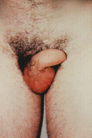
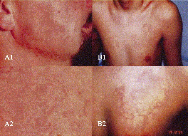
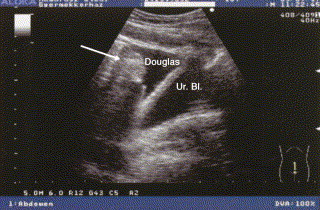
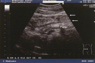
Hereditary angioedema (HAE), a rare but life-threatening condition, manifests as acute attacks of facial, laryngeal, genital, or peripheral swelling or abdominal pain secondary to intra-abdominal edema. Resulting from mutations affecting C1 esterase inhibitor (C1-INH), inhibitor of the first complement system component, attacks are not histamine-mediated and do not respond to antihistamines or corticosteroids. Low awareness and resemblance to other disorders often delay diagnosis; despite availability of C1-INH replacement in some countries, no approved, safe acute attack therapy exists in the United States. The biennial C1 Esterase Inhibitor Deficiency Workshops resulted from a European initiative for better knowledge and treatment of HAE and related diseases. This supplement contains work presented at the third workshop and expanded content toward a definitive picture of angioedema in the absence of allergy. Most notably, it includes cumulative genetic investigations; multinational laboratory diagnosis recommendations; current pathogenesis hypotheses; suggested prophylaxis and acute attack treatment, including home treatment; future treatment options; and analysis of patient subpopulations, including pediatric patients and patients whose angioedema worsened during pregnancy or hormone administration. Causes and management of acquired angioedema and a new type of angioedema with normal C1-INH are also discussed. Collaborative patient and physician efforts, crucial in rare diseases, are emphasized. This supplement seeks to raise awareness and aid diagnosis of HAE, optimize treatment for all patients, and provide a platform for further research in this rare, partially understood disorder.


Figures

Fig 1
Fig 1
Facial edema. Photo: Dermatologische Klinik, Universitätsspital Zurich, Switzerland. Brunello Wüthrich, MD. Reprinted with permission from Swiss Medical Weekly.
Fig 2
Fig 2
Penile edema. Photo credit: Dr. Martin Ludovic.
Fig 3
Fig 3
Various erythematous rashes preceding or accompanying angioedema episodes. A1, Facial erythema marginatum; A2, close view. Photo credit: Brunello Wüthrich, MD. Reprinted with permission from Swiss Medical Weekly.B1, Mottling on chest; B2, close view. Photo credit: George Harmat, MD. Reprinted with permission from Acta Dermato-Venereologica.
Fig 4
Fig 4
Sagittal sonogram during an abdominal HAE attack. A significant amount of fluid can be seen in the pouch of Douglas, with a swollen intestinal loop visible (arrow) floating in the free fluid.
Fig 5
Fig 5
Transverse sonogram during an abdominal HAE attack, showing bowel and pancreas. Longitudinal section of a swollen bowel: the intestinal wall is edematously thickened (arrows); in addition, the reflectivity of the pancreas is increased.
Fig 6
Fig 6
Sonogram during an abdominal attack of HAE, showing kidney and spleen. Section of a thickened intestinal wall (large arrow) and a small amount of fluid (small arrow) between the left kidney and the spleen.
Fig 7
Fig 7
Sagittal and transverse sonograms during an HAE attack before and after treatment. Sagittal sections are shown above, transverse below. A, A large amount of free peritoneal fluid has accumulated in the pouch of Douglas and, in the sagittal section, a floating intestinal loop is visible. The urinary bladder appears below. B, Soon after treatment with C1-INH concentrate, the amount of peritoneal fluid is somewhat decreased. C, Only a minimal amount of fluid is present in the pouch of Douglas 24 hours after C1-INH treatment. Several sonograms have previously been published in slightly different format., Transverse panels B and C reprinted with permission from Acta Paediatrica. Sagittal panels A-C reprinted with permission from the European Journal of Gastroenterology and Hepatology.
Fig 8
Fig 8
Transverse sonogram during an abdominal HAE attack: liver and pancreas. Increased hepatic reflection (starry sky liver) and thickened, echogenic portal veins (arrow); the pancreatic region is also hyperechoic (double arrows). Reprinted with permission from Acta Paediatrica.
Fig 9
Fig 9
Transverse sonogram during an abdominal HAE attack: liver and cholecyst. Because of edematous swelling, the wall of the cholecyst is hyperechoic (large arrow) and the small portal veins are also more echogenic (small arrows) in contrast with the liver's overall decrease in echogenicity.
Fig 10
Fig 10
Structure of C1NH gene., Arrowheads represent Alu elements. The positions of the 4 polymorphic sites are indicated by elongated arrows. Nucleotide numbers refer to the C1NH gene, with number 1 denoting the first nucleotide of exon 1. Accession number X54486.
Fig 11
Fig 11
Mutation analysis of the C1NH gene by DHPLC showing data with insertion, point mutation, and deletion. Red and black profiles correspond to patient and control samples, respectively. A, A 10-nucleotide insertion in exon 3 causes the presence of heteroduplexes before and after normal homoduplex. B, A point mutation (A → G) in exon 4 causes the presence of heteroduplexes in close vicinity to a normal homoduplex. C, A 5-nucleotide deletion in exon 5 causes the presence of heteroduplexes before normal homoduplex. Drouet et al, Unpublished data, August 2003.
Fig 12
Fig 12
Analysis of C1NH gene mutations by SSCA. A, SSCA of fragment 2144-2360 including part of exon 3; lanes 1, 2, 4 HAE kindreds without mutations in this fragment; lane 3, mutation 2264-5delAG [R18fsX33]. B, SSCA of fragment 2482-2761 including part of exon 3; lane 1, healthy control; lanes 3, 4, HAE kindreds without mutations in this fragment; lane 2, mutation in exon 3: T2650C [F147S]. C, SSCA of fragment 8690-8895 including exon 6; lanes 1, 3, 4, HAE kindreds without mutations in exon 6; lane 2, exon 6 mutation: C8823G [Y308Stop]. Lopez-Trascasa et al, Unpublished data, June 2003.
Fig 13
Fig 13
Southern blot analysis of patients with HAE-I carrying rearrangements in the C1NH gene. The outer lanes (N) contain control DNA of unaffected individuals and flank different heterozygous deletions/duplications. BclI-digested DNA was hybridized with a full-length cDNA probe. The 20-kb BclI fragment contains the 8 exons of the C1NH gene. Data from Stoppa-Lyonnet et al.
Fig 14
Fig 14
Quantitative fluorescence multiplex analysis: exon-specific multiplex PCR results for exons 3, 4, 5, 7, and 8 from C1NH and a control exon from BRCA1. Red and blue profiles correspond to the patient and control samples, respectively. Deletions of exons 3 and 4 (A), duplication of exon 4 (B), deletion of exon 8 (C), and deletion of exon 4 (D) are clearly visible. ex, Exon. Modified from Duponchel et al.
Fig 15
Fig 15
Decision algorithm for mutation analyses of the C1NH gene. This algorithm takes into account the importance of mutations in the coding sequence and in the introns, and the abundance of rearrangements. Depending on the methods used, a preliminary screening of rearrangements may be preferred.
Fig 16
Fig 16
α-1-Antitrypsin tertiary structure conserved among the serpin family α-1-antitrypsin is composed of 3 β-sheets (A in red, B in green, and C in yellow) and 9 α-helices (A-I in gray). The blue strand is the reactive center loop (RCL) with the P1 active amino acid. Reprinted with permission from Silverman et al.
Fig 17
Fig 17
Different pathways of kinin generation in C1-inhibitor deficiency state. Low C1-inhibitor leads to uncontrolled activation of factor XII, which generates kallikrein and plasmin. Kallikrein liberates bradykinin from HK, whereas plasmin cleaves off C2-kinin from activated C2. Activated C2 is continuously produced during baseline complement activation, which is increased as a result of insufficient control of autoactivation of C1 caused by C1-INH deficiency.
Fig 18
Fig 18
Analysis of vascular permeability. Extravasation of Evans blue dye at 15 to 30 minutes was much more extensive in C1-INH–deficient mice (B) than in wild-type mice (A), particularly after the application of mustard oil to the left ears of the mice. (C) The difference in the amount of extravasation was clearly demonstrated by the rear footpads of mice of each genotype. Administration of human C1-INH resulted in reduced vascular permeability, as did the combination of Bk2R deficiency together with C1-INH deficiency. Reprinted with permission of the American Society for Clinical Investigation.
Fig 19
Fig 19
Spectrophotometric analysis of vascular permeability in the small intestine. Quantitation of extravasated Evans blue dye in C1INH−/− mice treated with an ACE inhibitor (captopril), a Bk1R antagonist (des-Arg9,[Leu8]-bradykinin; Bk1RA), and a Bk2R antagonist (Hoe140). Reprinted with permission of the American Society for Clinical Investigation.
Fig 20
Fig 20
Spectrophotometric analysis of vascular permeability in footpads. Quantitation of Evans blue dye extracted from the paws of mice of the indicated genotypes, either treated or not treated with intravenous C1-INH (C1I), DX-88 (DX), or Hoe140 (Hoe). Reprinted with permission of the American Society for Clinical Investigation.
Fig 21
Fig 21
Pathways for bradykinin generation and metabolism. On the endothelium, bradykinin generation likely initiates independent of factor XII; however, once kallikrein is generated, it soon catalyzes factor XII activation, thus amplifying the response.
Fig 22
Fig 22
Correlation in female patients with HAE between serum progesterone (A) and SHBG concentrations (B) and the number of edematous attacks in the year after blood sampling. Spearman correlation coefficients and their significance (P values) are indicated.
Fig 23
Fig 23
Underlying angioedema disease in 44 women who had induction or exacerbation of recurrent angioedema caused by OCs or HRT.
Fig 24
Fig 24
Assessment of C4 antigen concentration by polyclonal anti-C4 antibodies. Assessment of C4 antigen levels by polyclonal anti-C4 antibodies might serve as a simple follow up parameter for therapy. Upper left and right panels depict C1-INH versus C4 antigen concentrations in 111 plasma samples from 21 patients with HAE-I. In the left panel, the individual C1-INH concentrations are plotted against C4 concentrations in plasma samples from patients with various therapeutic managements. Open square, no therapy; inversed open triangle, therapy with tranexamic acid; closed circle, therapy with danazol; closed triangle, therapy with stanozolol. Open squares and triangles represent samples from patients with current or recent bacterial or viral infections and therapy with danazol or stanozolol. Open squares with a crossing line indicate samples from an asymptomatic woman, a member of a family with concomitant heterozygous C1-INH deficiency and factor I protein gene mutation, herself heterozygous for C1-INH only. In the right panel, the means (±1 SD) of concentration ranges are depicted. The lower left panel indicates clinical manifestation of the C1-INH deficiency in 111 patients with HAE-I: closed circles give C1-INH concentration in plasma collected after an attack, and open circles indicate mean C1-INH concentrations of 2 assessments between which an attack occurred.
Fig 25
Fig 25
Work-up flow sheet for patients with a clinical history of idiopathic or unknown origin abdominal symptoms or angioedema.
Fig 26
Fig 26
Adverse events attributable to HAE or AAE in patients with and without a supply of C1 inhibitor at home.
Fig 27
Fig 27
Effect of long-term danazol treatment on serum concentrations of C4 (A) and C1-INH (B) in children with HAE. Baseline values are compared with those measured in the first serum sample obtained after treatment had been initiated. C4 levels were determined in 8 children. Patients with high concentrations of nonfunctional C1-INH (233, 214, and 297%) were excluded from the comparison. Danazol treatment significantly increased both C4 and C1-INH levels (P = .002 and P = .0156, respectively; Wilcoxon signed-rank test).
Fig 28
Fig 28
Flow of data in the European HAE Register. PC, Personal computer.
Fig 29
Fig 29
Window for accessing the European HAE Register.
Fig 30
Fig 30
European HAE Register visit form.
Fig 31
Fig 31
Hungarian HAE Center patient accrual.
Fig 32
Fig 32
Hungarian HAE Center: flow of responsibilities.

Similar articles

Cited by

  • HAE Pathophysiology and Underlying Mechanisms.
    Zuraw BL, Christiansen SC. Zuraw BL, et al. Clin Rev Allergy Immunol. 2016 Oct;51(2):216-29. doi: 10.1007/s12016-016-8561-8. Clin Rev Allergy Immunol. 2016. PMID: 27459852 Review.
  • A nationwide survey of hereditary angioedema due to C1 inhibitor deficiency in Italy.
    Zanichelli A, Arcoleo F, Barca MP, Borrelli P, Bova M, Cancian M, Cicardi M, Cillari E, De Carolis C, De Pasquale T, Del Corso I, Di Rocco PC, Guarino MD, Massaro I, Minale P, Montinaro V, Neri S, Perricone R, Pucci S, Quattrocchi P, Rossi O, Triggiani M, Zanierato G, Zoli A. Zanichelli A, et al. Orphanet J Rare Dis. 2015 Feb 6;10:11. doi: 10.1186/s13023-015-0233-x. Orphanet J Rare Dis. 2015. PMID: 25758562 Free PMC article.
  • Throat microbiota alterations in patients with hereditary angioedema.
    Wang X, Cao Y, Zhi Y. Wang X, et al. World Allergy Organ J. 2022 Sep 28;15(10):100694. doi: 10.1016/j.waojou.2022.100694. eCollection 2022 Oct. World Allergy Organ J. 2022. PMID: 36254183 Free PMC article.
  • Genetics of Hereditary Angioedema Revisited.
    Germenis AE, Speletas M. Germenis AE, et al. Clin Rev Allergy Immunol. 2016 Oct;51(2):170-82. doi: 10.1007/s12016-016-8543-x. Clin Rev Allergy Immunol. 2016. PMID: 27116602 Review.
  • C1 inhibitor: molecular and clinical aspects.
    Cicardi M, Zingale L, Zanichelli A, Pappalardo E, Cicardi B. Cicardi M, et al. Springer Semin Immunopathol. 2005 Nov;27(3):286-98. doi: 10.1007/s00281-005-0001-4. Epub 2005 Nov 11. Springer Semin Immunopathol. 2005. PMID: 16267649 Review.

References

    1. Dennehy J.J. Hereditary angioneurotic edema: report of a large kindred with defect in C'1 esterase inhibitor and review of the literature. Ann Intern Med. 1970;73:55–59. - PubMed
    1. Hawthorne N. Random House; New York: 2001. The house of the seven gables.
    1. Milton I.L. On giant urticaria. Edinburgh Med J. 1876;22:513–514. - PMC - PubMed
    1. Quincke H.I. Über akutes umschriebenes Hautödem [About an acute described skin edema] Monatshe Prakt Dermatol. 1882;1:129–131.
    1. Osler W. Hereditary angio-neurotic oedema. Am J Med Sci. 1888;95:362–367. - PubMed

Publication types

MeSH terms