. 2004 Jun;5(2):111-25.
doi: 10.1007/s10162-003-4012-3.

Postsynaptic targets of type II auditory nerve fibers in the cochlear nucleus

Affiliations

Postsynaptic targets of type II auditory nerve fibers in the cochlear nucleus

Thane E Benson et al. J Assoc Res Otolaryngol. 2004 Jun.

Abstract

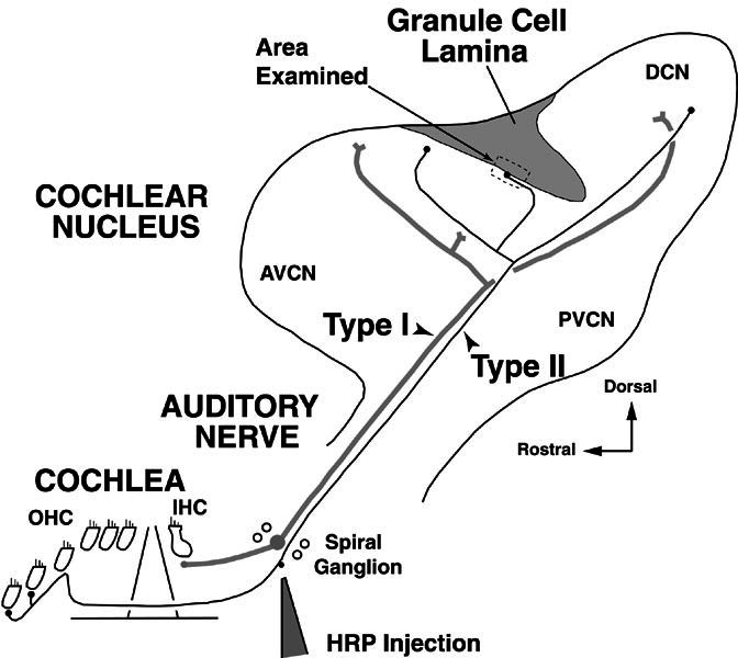
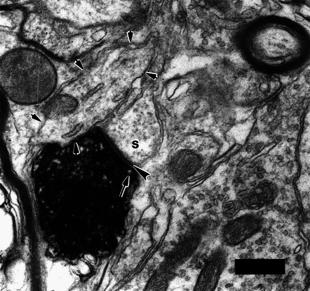
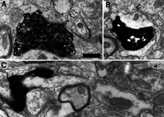
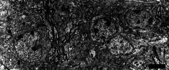
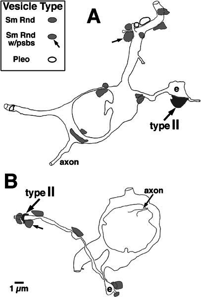
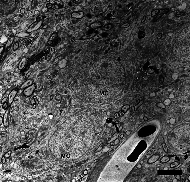
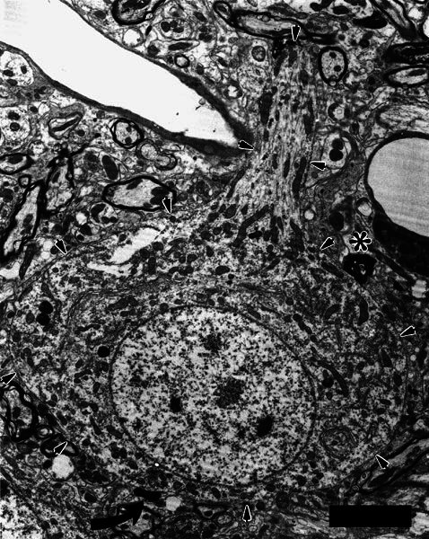
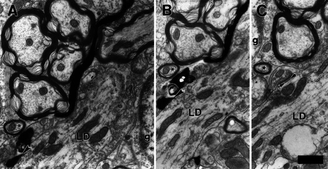
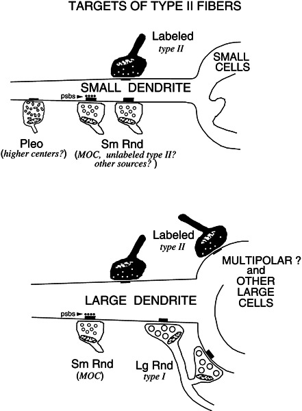
Type II auditory nerve fibers, which provide the primary afferent innervation of outer hair cells of the cochlea, project thin fibers centrally and form synapses in the cochlear nucleus. We investigated the postsynaptic targets of these synapses, which are unknown. Using serial-section electron microscopy of fibers labeled with horseradish peroxidase, we examined the border of the granule-cell lamina in mice, an area of type II termination that receives branches having swellings with complex shapes. About 70% of the swellings examined with the electron microscope formed morphological synapses, which is a much higher value than found in previous studies of type II swellings in other parts of the cochlear nucleus. The high percentage of synapses enabled a number of postsynaptic targets to be identified. Most of the targets were small dendrites. Two of these dendrites were traced to their somata of origin, which were cochlear-nucleus "small cells" situated at the border of the granule-cell lamina. These cells did not appear to receive any terminals containing synaptic vesicles that were large and round, indicating a lack of input from type I auditory nerve fibers. Nor did type II swellings or targets participate in the synaptic glomeruli formed by mossy terminals and the dendrites of granule cells. Other type II synapses were axosomatic and their targets were large cells, which were presumed multipolar cells and one cell with characteristics of a globular bushy cell. These large cells almost certainly receive additional input from type I auditory nerve fibers, which provide the afferent innervation of the cochlear inner hair cells. A few type II postsynaptic targets-the two small cells as well as a large dendrite-received synapses that had accompanying postsynaptic bodies, a likely marker for synapses of medial olivocochlear branches. These targets thus probably receive convergent input from type II fibers and medial olivocochlear branches. The diverse nature of the type II targets and the examples of segregated convergence of other inputs illustrates the synaptic complexity of type II input to the cochlear nucleus.


Figures

Figure 1
Figure 1
Schematic showing the site of horseradish peroxidase (HRP) injection to label type II fibers in the spiral ganglion of the cochlea. Auditory nerve fibers are shown peripherally in the cochlea where type II fibers form the afferent innervation of the outer hair cells (OHC) and type I fibers form the afferent innervation of the inner hair cells (IHC). Both fiber types project centrally in the auditory nerve, bifurcate in the cochlear nucleus, and form branches in the anteroventral, posteroventral, and dorsal subdivisions of the cochlear nucleus (AVCN, PVCN, and DCN). Dividing the VCN from the DCN is the granule-cell lamina, a region of termination for many type II fibers. Type II fibers in the lamina were examined in the approximate location indicated by the dashed box. The figure orientation is approximately the sagittal plane (see compass indicating dorsal toward the top of the figure). In successive figures, the orientation is reversed (dorsal toward the bottom of the figure) in order to be consistent with our previous work (Brown et al. ; Benson and Brown ; Benson et al. 1996).
Figure 2
Figure 2
Electron micrograph of a labeled type II terminal swelling forming a synapse on a dendritic spine (s). The specialization demonstrates the morphological criteria for synapses: synaptic vesicles (one denoted by arrow); synaptic cleft and postsynaptic density (large arrowhead). In this example the density is darker and thicker than in most other type II synapses (Fig. 3). The dense HRP reaction product typically obscures most morphology in the swelling. The dendritic spine was connected in other sections of the series to a “small dendrite” (outlined by small arrows; also illustrated in Fig. 3A). Scale bar = 0.5 μm. The scale bar is positioned upon an unlabeled terminal that contains pleomorphic vesicles.
Figure 3
Figure 3
Electron micrographs of three small dendrites (delineated with small arrowheads) receiving synapses from type II fibers. In panels A and C the postsynaptic density is indicated with large arrowheads, but in panel B the labeled terminal was synaptic in other sections. In panel A, the small dendrite could not be traced to its soma of origin, but it received a second synapse from the same labeled terminal onto a spine in a nearby section (Fig. 2). The small dendrite of panel B became much larger in nearby sections, as if swollen relative to other parts of the dendrite. This dendrite was followed through serial sections to the soma of a small cell (Figs. 4, 5A). The small dendrite in panel C is sectioned relatively longitudinally and is seemingly engulfed by the labeled type II terminal, a swelling found at a branch point of the fiber. The dendrite in C was also connected, through serial sections, to a small cell (Fig. 5B). Arrow denotes density of a synapse formed by an unlabeled terminal containing small round vesicles. Scale bar in A = 0.5 μm and also applies to B and C.
Figure 4
Figure 4
Electron micrograph of a small cell (SC) and a nearby cluster of granule cells (g). The small cell received, on its small dendrite, a synapse from an en passant swelling of a type II axon (Figs. 3B, 5A). Like granule cells, the small cell has chromatin aggregates in its nucleus, but it is larger than the granule cells and has a distinctive nuclear fold. Its perikaryal organelles are more densely packed, thus its cytoplasm appears darker than that of the granule cells. Scale bar = 5 μm.
Figure 5
Figure 5
Silhouettes of two small cells that received synapses from type II fibers onto their small dendrites. The dark shading shows the positions of the labeled type II terminals that formed the synapses (synapse for the cell of panel A is shown in Fig. 3B; synapse for the cell of panel B is shown in Fig. 3C). Also indicated are the positions of other, unlabeled terminals that formed synapses onto these small cells, coded according to the shape of their synaptic vesicles (Sm Rnd: small and round; Pleo: pleomorphic) (Schwartz and Gulley ; Cant and Morest ; Benson et al. 1996), as well as the presence of postsynaptic bodies (Sm Rnd w/psbs) in the postsynaptic element. The approximate sizes of these terminals on the figure are taken as the maximal silhouette area observed in the series of sections through the terminals. Each cell received synaptic input onto a dendritic enlargement (e). The cell in panel A was pictured in Figure 4.
Figure 6
Figure 6
Two presumed multipolar cells (MC) that received axosomatic synapses from labeled type II terminals. Swellings of the labeled type II fiber are indicated by two black, curved arrows. The swelling at the lower middle of the micrograph is a terminal swelling of the type II fiber and the swelling at the upper middle of the micrograph is a branch point swelling of the same type II fiber. This latter swelling synapses with a bootlike somatic appendage of the upper cell (outlined by small arrows). The cells are on the border of the lamina; a granule cell (g) is visible at lower right. Also at lower right, curving under the granule cell, is another labeled type II fiber (small arrows) illustrating the typical thin diameter of these fibers. Thicker-labeled myelinated fibers (m) and terminals from type I fibers within the core of the cochlear nucleus are visible at upper left. Scale bar = 5 μm.
Figure 7
Figure 7
Electron micrograph of a large neuron (outlined by arrows) that received two axosomatic synapses from a terminal swelling of a type II fiber. The neuron was located at the border of the granule-cell lamina, and it gave off a large dendrite directed toward the core of the ventral cochlear nucleus. This dendrite receives several terminals, one of which is darkened with HRP label. Another labeled axosomatic terminal is visible just beneath the asterisk. The origin of that terminal could not be determined unambiguously, but it connected with other axosomatic terminals that also formed punctate synapses with the neuron. Together, the terminals comprise a modified endbulb, suggesting that this neuron is a globular bushy cell that receives input from both type I and type II auditory nerve fibers. Scale bar = 5 μm.
Figure 8
Figure 8
Large dendrite (LD) that received a synapse (arrowheads in panels A and B) from a type II axon. The dendrite could not be traced to its soma, but its large diameter makes it distinct from the small dendrites of Figure 3. C The same large dendrite, in another section, also receives an unlabeled synapse with a robust postsynaptic density and with four postsynaptic bodies (between arrows), which is a synapse typical of those formed by medial olivocochlear branches (see text). Part of a granule cell (g) is indicated. In all panels, the asterisk denotes an axon with a relatively constant orientation relative to the large dendrite. Scale bar = 1 μm.
Figure 9
Figure 9
Summary of the synaptic targets of type II fibers observed in the present study. Most of the targets are small dendrites, at least some of which emanate from cochlear-nucleus small cells. Some targets are cell bodies of presumed multipolar cells. Occasional targets were large dendrites and presumed bushy cells. On the figure, labeled type II synaptic terminals are shaded in black and unlabeled synaptic terminals are unshaded. The unlabeled terminals are classified by the size and shape of their synaptic vesicles (abbreviations as in Fig. 5). Their potential sources are listed in italics.

Similar articles

Cited by

References

    1. Benson TE, Brown MC. Synapses formed by olivocochlear axon branches in the mouse cochlear nucleus. J. Comp. Neurol. 1990;295:52–70. - PubMed
    1. Benson TE, Berglund AM, Brown MC. Synaptic input to cochlear-nucleus dendrites that receive medial olivocochlear synapses. J. Comp. Neurol. 1996;365:27–41. - PubMed
    1. Berglund AM, Brown MC. Central trajectories of type-II spiral ganglion cells from various cochlear regions in mice. Hear. Res. 1994;75:121–130. - PubMed
    1. Berglund AM, Benson TE, Brown MC. Synapses from labeled type II axons in the mouse cochlear nucleus. Hear. Res. 1996;94:31–46. - PubMed
    1. Brawer JR, Morest DK. Relations between auditory nerve endings and cell types in the cat’s anteroventral cochlear nucleus seen with the Golgi method and Nomarski optics. J. Comp. Neurol. 1975;160:491–506. - PubMed

Publication types

LinkOut - more resources