. 2004 Jun;5(2):126-43.
doi: 10.1007/s10162-003-4029-7.

Efferent actions in the chinchilla vestibular labyrinth

Affiliations

Efferent actions in the chinchilla vestibular labyrinth

Vladimir Marlinski et al. J Assoc Res Otolaryngol. 2004 Jun.

Abstract

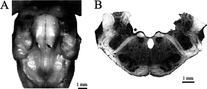
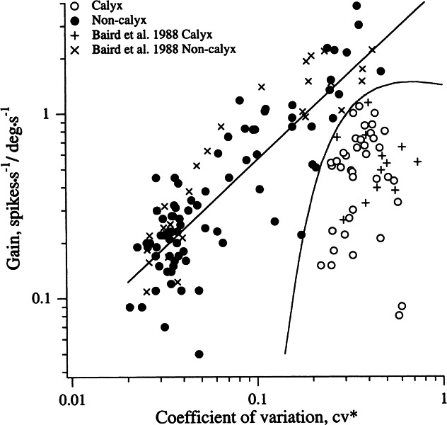
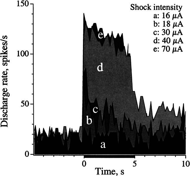
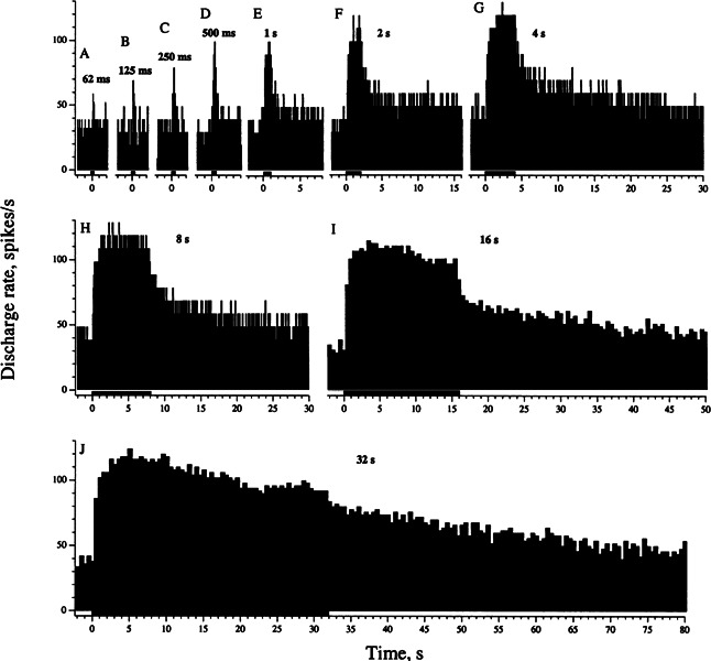
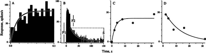
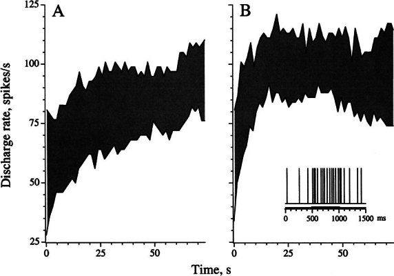
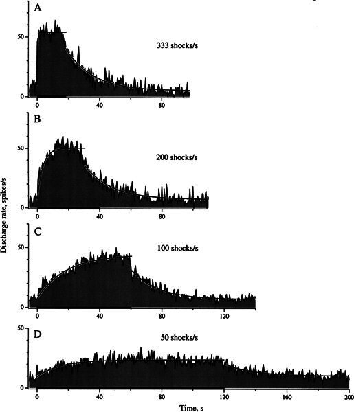
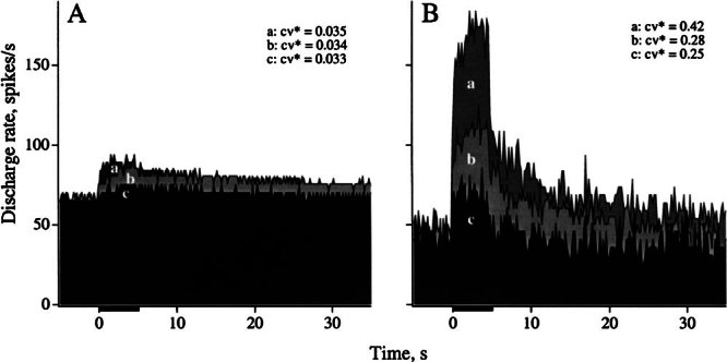
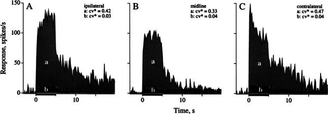
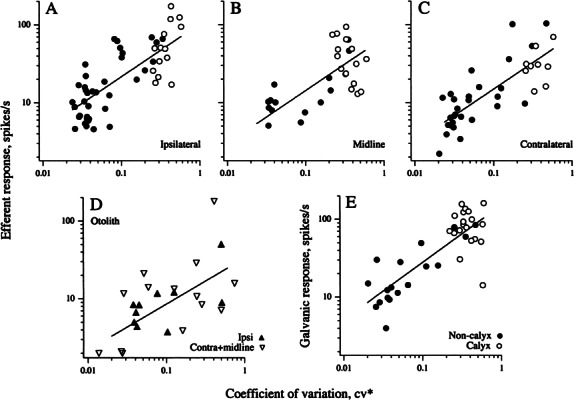
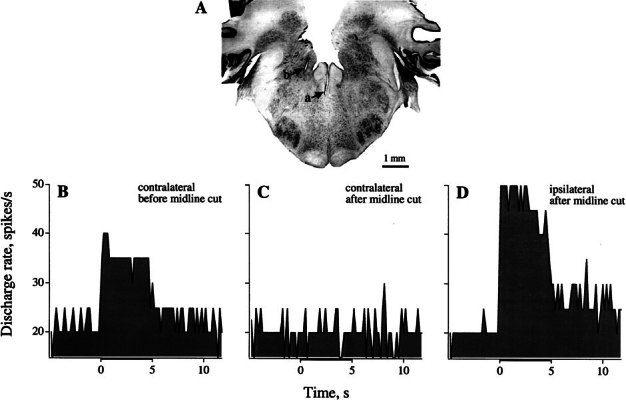
Efferent fibers were electrically stimulated in the brain stem, while afferent activity was recorded from the superior vestibular nerve in barbiturate-anesthetized chinchillas. We concentrated on canal afferents, but otolith afferents were also studied. Among canal fibers, calyx afferents were recognized by their irregular discharge and low rotational gains. In separate experiments, stimulating electrodes were placed in the efferent cell groups ipsilateral or contralateral to the recording electrode or in the midline. While single shocks were ineffective, repetitive shock trains invariably led to increases in afferent discharge rate. Such excitatory responses consisted of fast and slow components. Fast components were large only at high shock frequencies (200-333/s), built up with exponential time constants <0.1 s, and showed response declines or adaptation during shock trains >1 s in duration. Slow responses were obtained even at shock rates of 50/s, built up and decayed with time constants of 15-30 s, and could show little adaptation. The more regular the discharge, the larger was the efferent response of an afferent fiber. Response magnitude was proportional to cv*b, a normalized coefficient of interspike-interval variation (cv*) raised to the power b = 0.7. The value of the exponent b did not depend on unit type (calyx vs. bouton plus dimorphic, canal vs. otolith) or on stimulation site (ipsilateral, contralateral, or midline). Responses were slightly smaller with contralateral or midline stimulation than with ipsilateral stimulation, and they were smaller for otolith, as compared to canal, fibers. An anatomical study had suggested that responses to contralateral afferent stimulation should be small or nonexistent in irregular canal fibers. The suggestion was not confirmed in this study. Contralateral responses, including the large responses typically seen in irregular fibers, were abolished by shallow midline incisions that should have severed crossing efferent axons.


Figures

Figure 1
Figure 1
A Photograph of the brain stem and fourth ventricle in an animal with a midline placement of stimulating electrodes. Locations of the four electrodes, including the most effective one (arrow), are seen. B A cross-section from the same brain with an electrolytic lesion made with the most effective stimulating electrode. In each panel, a star marks the groove of the sulcus limitans, which serves as a landmark since it is just dorsal to the efferent cell group.
Figure 2
Figure 2
Semicircular canal afferents can be categorized as calyx or noncalyx units by their discharge properties. Shown is the relation between the gain in response to 2-Hz, 20-deg/s sinusoidal rotations and discharge regularity, the latter measured by a normalized coefficient of variation (cv*). Labeled calyx (+) and noncalyx (x) afferents from Baird et al. (1998) were used to construct the curved line, a quadratic discriminant function, which was then used to classify the unlabeled units of the present study as calyx (○) or noncalyx (●). Gains of noncalyx units related to cv* by a power law (straight line).
Figure 3
Figure 3
Responses of a noncalyx HC unit (cv* = 0.17) to electrical stimulation of the contralateral brain stem, 5-s shock trains (horizontal bar), 333 shocks/s. Shock intensity was varied from near-threshold (16 μA) to suprathreshold values (see legend). Responses are plotted from a discharge rate of 17 spikes/s, the average background discharge of the unit.
Figure 4
Figure 4
Shock-train duration distinguished a fast and a slow response component. Responses of a calyx SC unit (cv* = 0.25) to electrical stimulation of the brain-stem midline, 333 shocks/s, 58 μA (3xT). Shock-train duration was varied from 62 ms to 32 s (horizontal bars). Histogram for 62 ms, averaged for 3 repetitions; other histograms, one repetition. Bins: 0.1 s (62 ms–8 s), 0.5 s (16 and 32 s).
Figure 5
Figure 5
Kinetics of fast and slow response components. Same calyx unit as in Fig. 4. A Rate increase during the first 500 ms of the shock train illustrates the buildup of the fast response, which is fit by an exponential with a 80-ms time constant. An average of 7 repetitions. B Response to 16-s shock train. The decay of the slow response, starting 1 s into the post-train period, is fit by an exponential. The difference in the abrupt changes in rate at the beginning (F1) and end (F2) of the shock train reflects the adaptation of the fast response. The amplitude of the slow response (S) is estimated by the extrapolation of the exponential fit back to the start of the post-train period. F1 is the response averaged during the first 0.5 s of the shock train and F2 is the difference in rates during the last 1 s of the shock train and the slow response amplitude (S). CS is plotted versus the duration of the preceding shock train and fit by a simple exponential with a time constant of 2.7 ± 0.9 s. DF2 is plotted as a function of train duration and fit by an exponential with a time constant of 12.3 ± 2.3 s.
Figure 6
Figure 6
Adaptation of slow and fast response components. Shock trains, 0.5-s duration, 333 shocks/s are repeated every 1.5 s. There are a total of 50 trains. Rates are calculated during each train (T1, dark bar in inset to B) and in the 0.5 s immediately preceding period (T0). The slow response builds up with successive trains. The fast response is measured as the difference in rates between T1 and the immediately preceding T0 (shaded areas). A A noncalyx HC unit (cv* = 0.16), ipsilateral stimulation. The slow response continues to increase during the paradigm, while the fast response declines from 52 to 34 spikes/s. B A calyx SC unit (cv* = 0.25), midline stimulation. The slow response declines from 57 to 39 spikes/s; the fast response declines from 47 to 40 spikes/s.
Figure 7
Figure 7
Fast and slow responses behave differently as shock frequency changes. Same unit as in Fig. 4. Shock frequencies: 333 (A), 200 (B), 100 (C), and 50/s (D); train duration increases so that the number of shocks remains at 6000. A fast response is seen only at the beginning of the higher-frequency shock train. A slow response is seen in the post-train period at all frequencies and is also observed uncontaminated by a fast response during low-frequency shock trains. Separate simple exponentials are fit to the per-train response buildup and post-train response decay.
Figure 8
Figure 8
Efferent responses are larger in irregular, as compared to regular units. Comparison of responses in three regular (A) and three irregular units (B). Ipsilateral stimulation, 5-s shock trains, 333/s in all cases. For each panel, units had similar background discharges: A 66.8 (a), 65.4 (b), 65.8 (c) spikes/s and B 41.2 (a), 49.8 (b), and 44.0 (c) spikes/s. All units were HC except Bb, an SC unit. Calyx units are Ba and Bc. Responses are plotted from an averaged discharge rate of 66 (A) and 45 spikes/s (B).
Figure 9
Figure 9
Efferent responses are similar for ipsilateral (A), midline (B), and contralateral (C) stimulation. Each panel shows responses for an irregular (a) and a regular unit (b). Irregular afferents were calyx units except Ca.
Figure 10
Figure 10
Efferent response magnitudes parallel responses to galvanic currents. For canal afferents responses averaged over a 5-s efferent shock train are plotted against a normalized coefficient of variation (cv*) for ipsilateral (A), midline (B), and contralateral (C) efferent stimulation. D Efferent response magnitude versus cv* for otolith afferents. Responses to ipsilateral stimulation are indicated by filled triangles; responses to midline and contralateral stimulation are indicated by inverted open triangles. A–D 333/s, 40–70 μA shocks. E Relation between the response to 50-μA galvanic currents aid cv* . Straight lines are power-law fits; calyx and noncalyx units indicated by unfilled (○) and filled symbols (●), respectively. Slopes of the fitted lines in A–D and E are similar.
Figure 11
Figure 11
Midline lesion abolishes response to contralateral efferent stimulation. Recording from a noncalyx, SC unit (cv* = 0.08); efferent stimulation, 333/s, 5 s, 50 μA. A Cross-section of brain stem shows midline lesion (a) and electrolytic mark of ipsilateral stimulation site (b). Response to contralateral stimulation (B) is abolished after midline lesion (C). The stimulating array was then moved to ipsilateral side and a large response was obtained (D).

Similar articles

Cited by

References

    1. Art JJ, Fettiplace R, Fuchs PA. Synaptic hyperpolarization and inhibition of turtle cochlear hair cells. J. Physiol. 1984;356:525–550. - PMC - PubMed
    1. Ashmore JF, Russell IJ. Effects of efferent nerve stimulation on hair cells of the frog sacculus. J. Physiol. 1982;329:25–26.
    1. Bailey GP, Sewell WF. Calcitonin gene-related peptide suppresses hair cell responses to mechanical stimulation in the Xenopus lateral line organ. J. Neurosci. 2000;20:5163–5169. - PMC - PubMed
    1. Baird RA, Desmadryl G, Fernández C, Goldberg JM. The vestibular nerve of the chinchilla. II. Relation between afferent response properties and peripheral innervation patterns in the semicircular canals. J. Neurophysiol. 1988;60:182–203. - PubMed
    1. Bernard C, Cochran SL, Precht W. Presynaptic actions of cholinergic agents upon the hair cell-afferent fiber synapses in the vestibular labyrinth of the frog. Brain Res. 1985;338:225–236. doi: 10.1016/0006-8993(85)90151-9. - DOI - PubMed

Publication types

LinkOut - more resources