Comparative Study
. 2004 Sep 9:4:21.
doi: 10.1186/1472-6750-4-21.

A high-throughput cell migration assay using scratch wound healing, a comparison of image-based readout methods

Affiliations
Comparative Study

A high-throughput cell migration assay using scratch wound healing, a comparison of image-based readout methods

Justin C Yarrow et al. BMC Biotechnol. .

Abstract

Background: Cell migration is a complex phenomenon that requires the coordination of numerous cellular processes. Investigation of cell migration and its underlying biology is of interest to basic scientists and those in search of therapeutics. Current migration assays for screening small molecules, siRNAs, or other perturbations are difficult to perform in parallel at the scale required to screen large libraries.

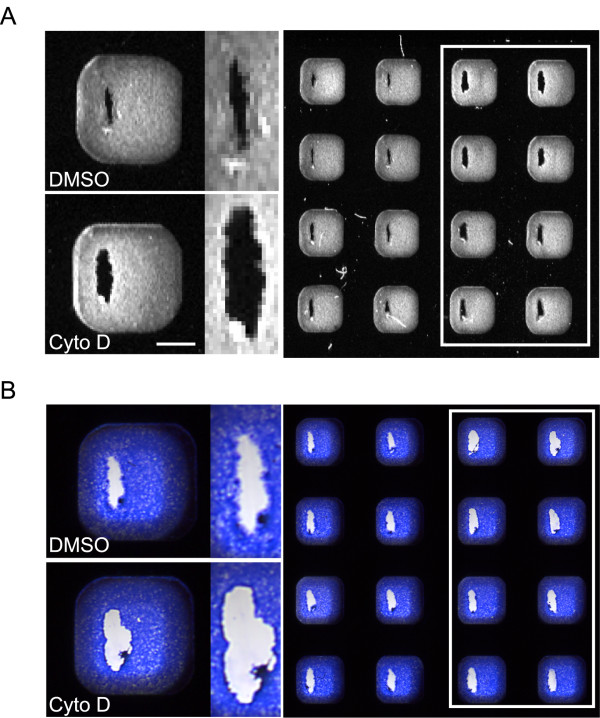
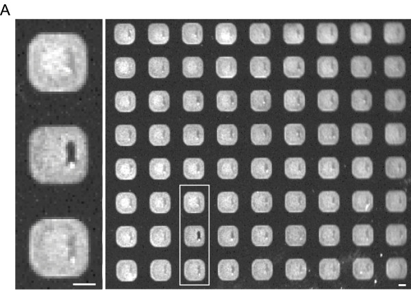
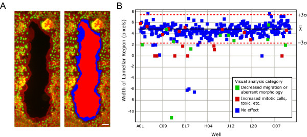
Results: We have adapted the commonly used scratch wound healing assay of tissue-culture cell monolayers to a 384 well plate format. By mechanically scratching the cell substrate with a pin array, we are able to create characteristically sized wounds in all wells of a 384 well plate. Imaging of the healing wounds with an automated fluorescence microscope allows us to distinguish perturbations that affect cell migration, morphology, and division. Readout requires ~1 hr per plate but is high in information content i.e. high content. We compare readouts using different imaging technologies, automated microscopy, scanners and a fluorescence macroscope, and evaluate the trade-off between information content and data acquisition rate.

Conclusions: The adaptation of a wound healing assay to a 384 well format facilitates the study of aspects of cell migration, tissue reorganization, cell division, and other processes that underlie wound healing. This assay allows greater than 10,000 perturbations to be screened per day with a quantitative, high-content readout, and can also be used to characterize small numbers of perturbations in detail.


Figures

Figure 1
Figure 1
Adaptation of a wound healing assay to a 384 well plate format. (A) Images from a time-lapse sequence of BS-C-1 cells migrating on a coverslip to heal a wound. Bar 20 μm. (B) Images of wounds 0, 3, 7, 12, and 24 hrs after wounding show characteristic protrusion of lamella at 3 hrs, migration by 7 hrs, and eventual healing of the wound at 24 hrs. Cells are stained for filamentous actin. Bar 100 μm. (C) Schematic of the protocol used for screening. The 96 well pin array (shown) produces wounds with consistent shape and placement within each well. Image of 25 wells stained for filamentous actin 7 hrs after wounding. Bar 500 μm. (D) The primary phenotypes observed with an automated microscope in a small molecule screen (4× objective, 7 hr healing). Wells often showed a combination of these phenotypes. Cells are stained for filamentous actin and DNA. The dotted yellow line notes the approximate edge of the initial wound (based on the high density of nuclei). Bar 100 μm.
Figure 2
Figure 2
The use of scanners to assay wound healing after 7 hrs recovery. (A) An image of actin-stained wells obtained using a fluorescence scanner. Normal healing wounds (left two columns) can be distinguished from wells treated with a titration of the actin cytoskeleton disrupter cytochalasin D (white box) by the width of the wound and staining at the wound margin. (B) An image of Coomassie-stained wells obtained using a transmitted-light scanner. Normal healing wounds have a more diffuse wound margin, denoting cell migration into the wound, than wells treated with cytochalasin D (white box). Bar 1 mm.
Figure 3
Figure 3
The use of a fluorescence macroscope to assay wound healing after 24 hrs recovery. Fluorescence macroscope image of wells stained for filamentous actin 24 hrs after wounding. A well showing reduced wound healing can be easily distinguished (middle well white box) from wells showing complete healing which are seen as light grey streaks. Bar 1 mm.
Figure 4
Figure 4
Automated analysis of wound healing images. (A) An image showing normal migration and the same image after automated analysis defines regions. Extracting characteristics of the lamellar region (blue) allows us to distinguish between the phenotypes that affect wound healing (Figure 1). Stains: red – filamentous actin; green – DNA. Bar 100 μm. (B) Automated and visual analysis are complementary approaches. Automatically generated lamellar width data is plotted for a 384 well plate. Compounds found to affect wound healing by visual analysis are shown in green, and wells that differed from the control in other ways (increased mitotics, a disrupted monolayer, etc.) are shown in red. Mean lamellar width (black line) and three standard deviations (red dashed lines) are plotted for control wells. Data collected from screening one 384 well plate of small molecules.

References

    1. Wong MK, Gotlieb AI. The reorganization of microfilaments, centrosomes, and microtubules during in vitro small wound reendothelialization. J Cell Biol. 1988;107:1777–1783. doi: 10.1083/jcb.107.5.1777. - DOI - PMC - PubMed
    1. Coomber BL, Gotlieb AI. In vitro endothelial wound repair. Interaction of cell migration and proliferation. Arteriosclerosis. 1990;10:215–222. - PubMed
    1. Zahm JM, Kaplan H, Herard AL, Doriot F, Pierrot D, Somelette P, Puchelle E. Cell migration and proliferation during the in vitro wound repair of the respiratory epithelium. Cell Motil Cytoskeleton. 1997;37:33–43. doi: 10.1002/(SICI)1097-0169(1997)37:1<33::AID-CM4>3.0.CO;2-I. - DOI - PubMed
    1. Lampugnani MG. Cell migration into a wounded area in vitro. Methods Mol Biol. 1999;96:177–182. doi: 10.1385/1-59259-258-9:177. - DOI - PubMed
    1. Lu KV, Jong KA, Rajasekaran AK, Cloughesy TF, Mischel PS. Upregulation of tissue inhibitor of metalloproteinases (TIMP)-2 promotes matrix metalloproteinase (MMP)-2 activation and cell invasion in a human glioblastoma cell line. Lab Invest. 2004;84:8–20. doi: 10.1038/sj.labinvest.3700003. - DOI - PubMed

Publication types