. 2004 Sep 21;101(38):13861-6.
doi: 10.1073/pnas.0405908101. Epub 2004 Sep 9.

Enhanced expression of transient receptor potential channels in idiopathic pulmonary arterial hypertension

Affiliations

Enhanced expression of transient receptor potential channels in idiopathic pulmonary arterial hypertension

Ying Yu et al. Proc Natl Acad Sci U S A. .

Abstract

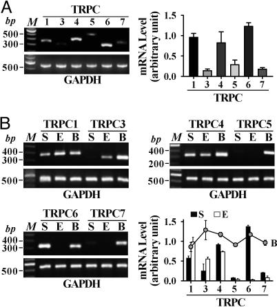
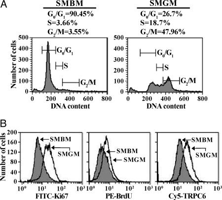
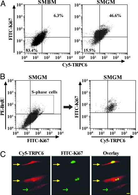
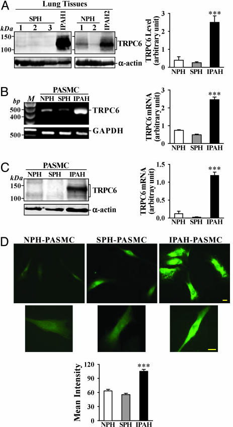
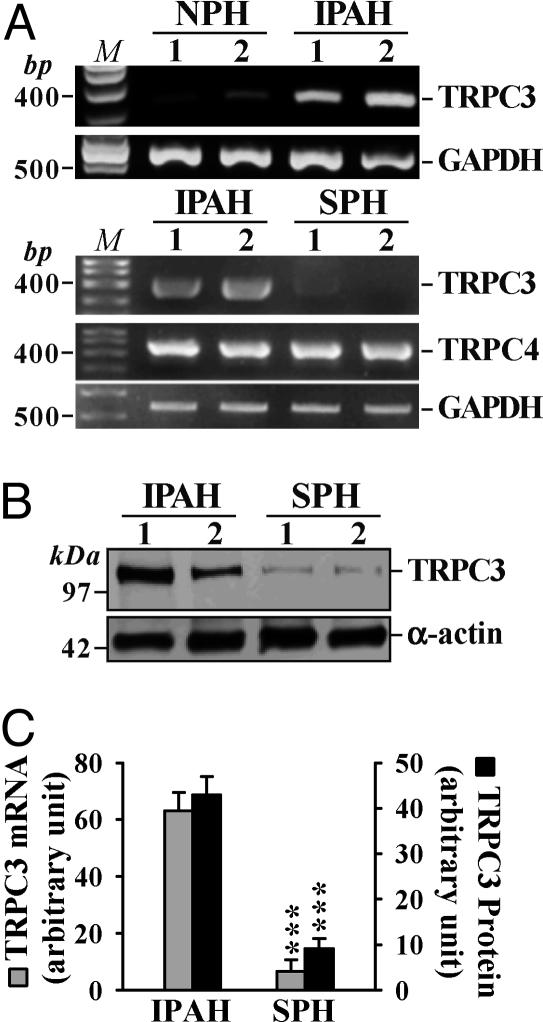
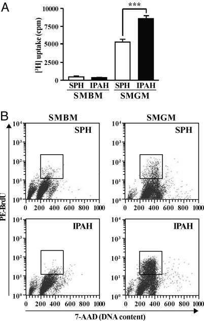
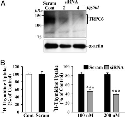
Pulmonary vascular medial hypertrophy caused by excessive pulmonary artery smooth muscle cell (PASMC) proliferation is a major cause for the elevated pulmonary vascular resistance in patients with idiopathic pulmonary arterial hypertension (IPAH). Increased Ca(2+) influx is an important stimulus for PASMC proliferation. Transient receptor potential (TRP) channel genes encode Ca(2+) channels that are responsible for Ca(2+) entry during cell proliferation. Normal human PASMC expressed multiple canonical TRP (TRPC) isoforms; TRPC6 was highly expressed and TRPC3 was minimally expressed. The protein expression of TRPC6 in normal PASMC closely correlated with the expression of Ki67, suggesting that TRPC6 expression is involved in the transition of PASMC from quiescent phase to mitosis. In lung tissues and PASMC from IPAH patients, the mRNA and protein expression of TRPC3 and -6 were much higher than in those from normotensive or secondary pulmonary hypertension patients. Inhibition of TRPC6 expression with TRPC6 small interfering RNA markedly attenuated IPAH-PASMC proliferation. These results demonstrate that expression of TRPC channels correlates with the progression of the cell cycle in PASMC. TRPC channel overexpression may be partially responsible for the increased PASMC proliferation and pulmonary vascular medial hypertrophy in IPAH patients.


Figures

Fig. 1.
Fig. 1.
The mRNA expression of TRPC channels in NPH lung tissue and PASMC. (A) Representative and summarized (n = 4) mRNA products for TRPC genes and GAPDH in NPH human lung. (B) Representative and summarized (n = 3) data showing TRPC gene mRNA products in human brain tissue (B), PASMC (S), and PAEC (E). M, 100-bp DNA ladder.
Fig. 2.
Fig. 2.
Cell cycle analysis of growth-arrested and proliferating PASMC and correlation of TRPC6 expression with PASMC proliferation. (A) Flow cytometry graphs (representative of three trials) of cell cycle analysis for growth-arrested (SMBM) and proliferating (SMGM) PASMC. Number of cells is plotted as a function of DNA content. (B) Graphs showing cell number as a function of Ki67 expression, BrdUrd incorporation, or TRPC6 expression in SMBM- and SMGM-treated PASMC.
Fig. 3.
Fig. 3.
Correlation of Ki67 expression and BrdUrd incorporation with TRPC6 expression. (A) Dot-plot analysis (representative of three trials) of TRPC6 expression vs. Ki67 expression in growth-arrested and proliferating PASMC. Upper right quadrants represent cells in the S phase. (B) Three-color dot-plot analysis of TRPC6 distribution vs. BrdUrd incorporation and Ki67 expression in proliferating PASMC (representative of three trials). The box in Left indicates the S-phase cells that are stained by both Ki67 and BrdUrd. (C) Fluorescent images (×40) showing PASMC stained by Ki67 and TRPC6. An overlay of the two fluorescent images identifies PASMC expressing both Ki67 and TRPC6. Yellow and green arrows indicate Ki67-positive and -negative PASMC, respectively. (Scale bar, 20 μm.)
Fig. 4.
Fig. 4.
TRPC6 mRNA and protein expression is increased in pulmonary tissues from IPAH patients. (A) Representative and summarized (NPH, n = 3; SPH, n = 8; IPAH, n = 3; normalized to α-actin) TRPC6 protein expression in SPH, NPH, and IPAH lung tissues and their α-actin controls. (B) Representative and summarized (n = 3 for each) data showing TRPC6 mRNA products in isolated NPH-, SPH-, or IPAH-PASMC. M, 100-bp DNA ladder. (C) Representative and summarized (n = 5 for each) TRPC6 protein expression in isolated PASMC from NPH, SPH, and IPAH patients. (D) Representative (Top, ×40; Middle, ×100) and summarized immunofluorescence staining of FITC-conjugated TRPC6 (Bottom) in isolated NPH-(n = 11), SPH (n = 21), and IPAH-(n = 36) PASMC. (Scale bars, 20 μm.) ***, P < 0.001 vs. NPH and/or SPH.
Fig. 5.
Fig. 5.
Up-regulated TRPC3 expression in IPAH-PASMC. (A) TRPC3 mRNA expression in PASMC from two NPH patients and two IPAH patients. TRPC3 and -4 mRNA expression in PASMC from two IPAH patients and two SPH patients. M, 100-bp DNA ladder. (B) TRPC3 protein expression in the same IPAH and SPH samples as shown in A. (C) Summarized bar graphs (n = 3 for each) depicting normalized TRPC3 mRNA and protein expression. ***, P < 0.001 vs. IPAH.
Fig. 6.
Fig. 6.
Increased proliferation of IPAH-PASMC. (A) [3H]Thymidine incorporation in SPH (n = 8) and IPAH (n = 8) PASMC before (12 h, SMBM) and 24 h after the addition of 5% FBS and growth factors (SMGM). ***, P < 0.001 vs. SMGM-SPH. (B) Bivariate distribution of BrdUrd vs. DNA content for SMBM- or SMGM-treated SPH- and IPAH-PASMC. Boxes indicate S phase PASMC. Data are representative of three experiments.
Fig. 7.
Fig. 7.
TRPC6 siRNA attenuates TRPC6 expression and proliferation in IPAH cells. (A) Effect of two concentrations of TRPC6-siRNA-2 expression vector, and its scrambled control (4 μg/ml), on TRPC6 protein expression. (B) (Left) Normalized [3H]thymidine uptake plotted for cells untreated (Cont) or treated with the scrambled siRNA duplex (Scram, 200 nM). (Right) Cell growth in cells transfected with two concentrations of either the scrambled (n = 8) or TRPC6 (n = 8) siRNA duplexes. Data are normalized to the basal [3H]thymidine uptake in the absence of siRNA (Cont). ***, P < 0.001 vs. Scram.

References

    1. Runo, J. R. & Loyd, J. E. (2003) Lancet 361, 1533–1544. - PubMed
    1. Fishman, A. P. (1998) Chest 114, 242S–247S. - PubMed
    1. Wagenvoort, C. A. (1960) Circulation 22, 535–546. - PubMed
    1. Wood, P. (1958) Br. Heart J. 20, 557–570. - PMC - PubMed
    1. Stenmark, K. R. & Mecham, R. P. (1997) Annu. Rev. Physiol. 59, 89–144. - PubMed

Publication types

MeSH terms