

Structure and DNA-binding properties of the cytolysin regulator CylR2 from Enterococcus faecalis
- PMID: 15359276
- PMCID: PMC517608
- DOI: 10.1038/sj.emboj.7600367
Structure and DNA-binding properties of the cytolysin regulator CylR2 from Enterococcus faecalis
Abstract
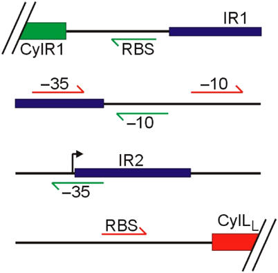
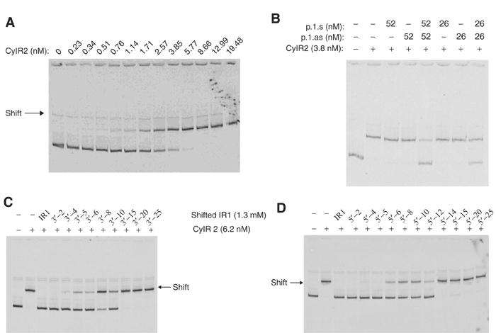
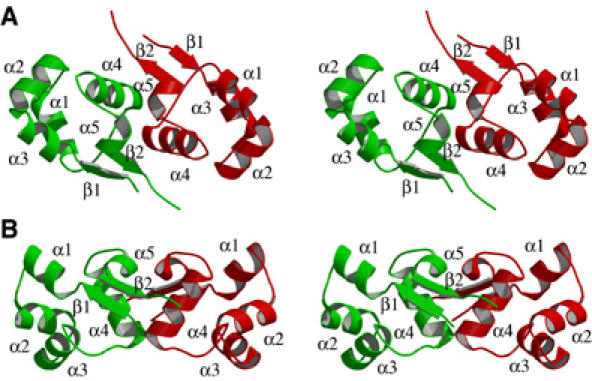
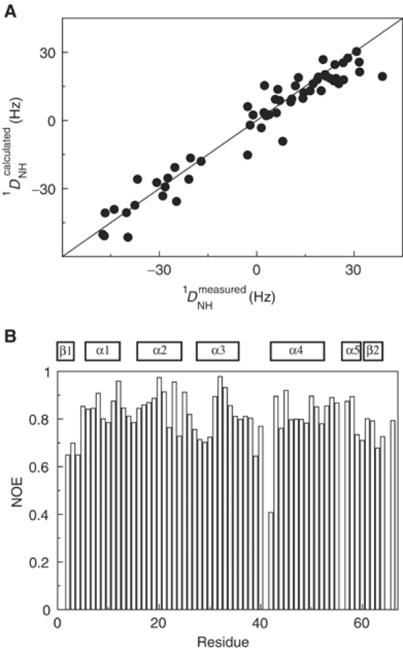
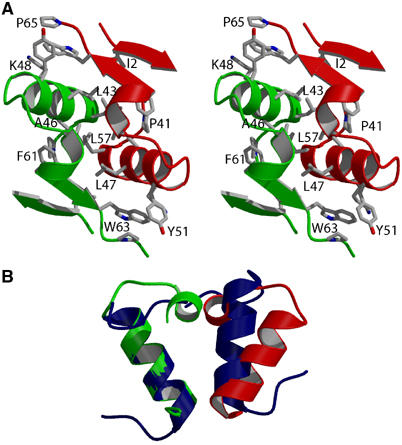
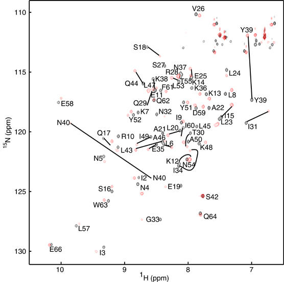
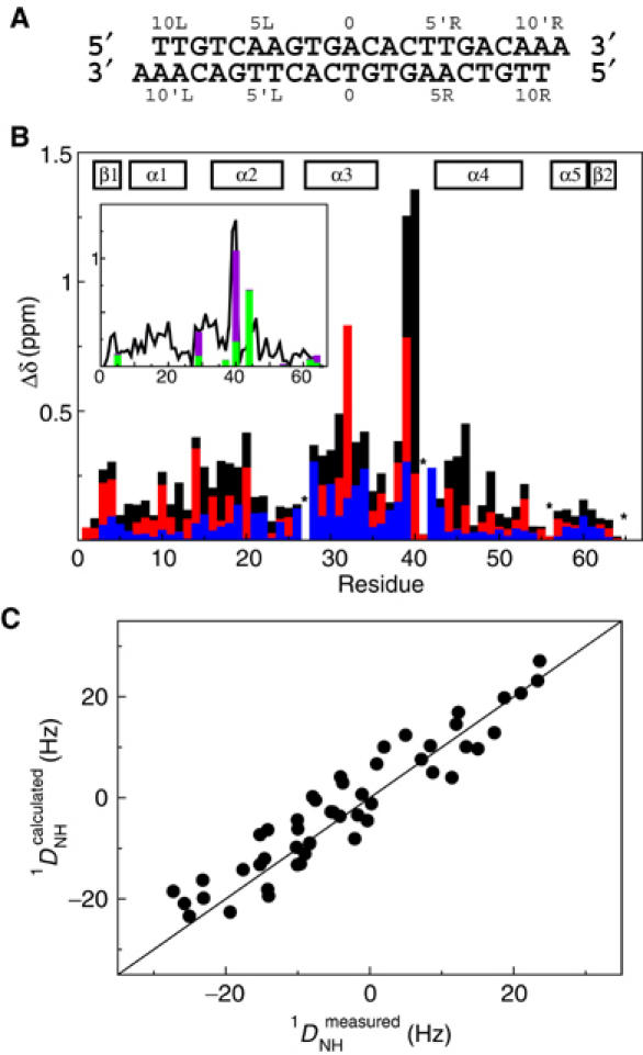
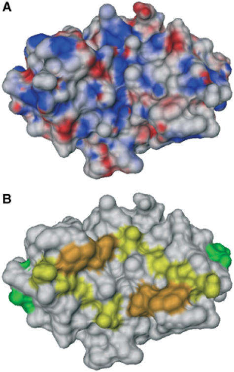
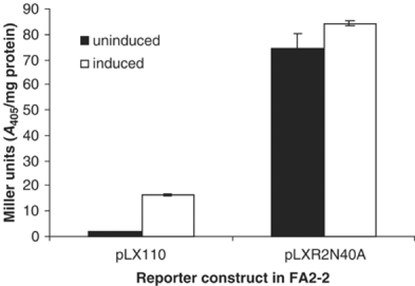
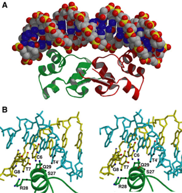
Enterococcus faecalis is one of the major causes for hospital-acquired antibiotic-resistant infections. It produces an exotoxin, called cytolysin, which is lethal for a wide range of Gram-positive bacteria and is toxic to higher organisms. Recently, the regulation of the cytolysin operon was connected to autoinduction by a quorum-sensing mechanism involving the CylR1/CylR2 two-component regulatory system. We report here the crystal structure of CylR2 and its properties in solution as determined by heteronuclear NMR spectroscopy. The structure reveals a rigid dimer containing a helix-turn-helix DNA-binding motif as part of a five-helix bundle that is extended by an antiparallel beta-sheet. We show that CylR2 is a DNA-binding protein that binds specifically to a 22 bp fragment of the cytolysin promoter region. NMR chemical shift perturbation experiments identify surfaces involved in DNA binding and are in agreement with a model for the CylR2/DNA complex that attributes binding specificity to a complex network of CylR2/DNA interactions. Our results propose a mechanism where repression is achieved by CylR2 obstruction of the promoter preventing biosynthesis of the cytolysin operon transcript.
Figures
Similar articles
-
Expression, purification, crystallization and preliminary crystallographic studies of the Enterococcus faecalis cytolysin repressor CylR2.Acta Crystallogr D Biol Crystallogr. 2004 Apr;60(Pt 4):746-8. doi: 10.1107/S0907444904002410. Epub 2004 Mar 23. Acta Crystallogr D Biol Crystallogr. 2004. PMID: 15039573
-
Two-component regulator of Enterococcus faecalis cytolysin responds to quorum-sensing autoinduction.Nature. 2002 Jan 3;415(6867):84-7. doi: 10.1038/415084a. Nature. 2002. PMID: 11780122
-
Enterococcal cytolysin: activities and association with other virulence traits in a pathogenicity island.Int J Med Microbiol. 2004 Apr;293(7-8):609-18. doi: 10.1078/1438-4221-00301. Int J Med Microbiol. 2004. PMID: 15149038 Review.
-
Crystallization and preliminary crystallographic analysis of an Enterococcus faecalis repressor protein, CylR2, involved in regulating cytolysin production through quorum-sensing.Acta Crystallogr D Biol Crystallogr. 2004 Jun;60(Pt 6):1145-8. doi: 10.1107/S0907444904008078. Epub 2004 May 21. Acta Crystallogr D Biol Crystallogr. 2004. PMID: 15159583
-
Structure, function, and biology of the Enterococcus faecalis cytolysin.Toxins (Basel). 2013 Apr 29;5(5):895-911. doi: 10.3390/toxins5050895. Toxins (Basel). 2013. PMID: 23628786 Free PMC article. Review.
Cited by
-
Bacterial cell-to-cell signaling in the gastrointestinal tract.Infect Immun. 2005 Jun;73(6):3197-209. doi: 10.1128/IAI.73.6.3197-3209.2005. Infect Immun. 2005. PMID: 15908344 Free PMC article. Review. No abstract available.
-
Combatting virulent gut bacteria by inhibiting the biosynthesis of a two-component lanthipeptide toxin.Nat Commun. 2025 Jul 28;16(1):6936. doi: 10.1038/s41467-025-62161-7. Nat Commun. 2025. PMID: 40721579 Free PMC article.
-
Transcriptional analysis of the Streptococcus pyogenes salivaricin locus.J Bacteriol. 2014 Feb;196(3):604-13. doi: 10.1128/JB.01009-13. Epub 2013 Nov 15. J Bacteriol. 2014. PMID: 24244008 Free PMC article.
-
Structural models of protein-DNA complexes based on interface prediction and docking.Curr Protein Pept Sci. 2011 Sep;12(6):531-9. doi: 10.2174/138920311796957694. Curr Protein Pept Sci. 2011. PMID: 21787304 Free PMC article.
-
Direct stimulus perception and transcription activation by a membrane-bound DNA binding protein.Mol Microbiol. 2009 Aug;73(3):482-91. doi: 10.1111/j.1365-2958.2009.06787.x. Epub 2009 Jul 7. Mol Microbiol. 2009. PMID: 19602149 Free PMC article.
References
-
- Aggarwal AK, Rodgers DW, Drottar M, Ptashne M, Harrison SC (1988) Recognition of a DNA operator by the repressor of phage 434: a view at high resolution. Science 242: 899–907 - PubMed
-
- Bax A, Grzesiek S (1993) Methodological advances in protein NMR. Accounts Chem Res 26: 131–138
-
- Bell AC, Koudelka GB (1993) Operator sequence context influences amino acid–base-pair interactions in 434 repressor–operator complexes. J Mol Biol 234: 542–553 - PubMed
-
- Booth MC, Bogie CP, Sahl HG, Siezen RJ, Hatter KL, Gilmore MS (1996) Structural analysis and proteolytic activation of Enterococcus faecalis cytolysin, a novel lantibiotic. Mol Microbiol 21: 1175–1184 - PubMed
Publication types
MeSH terms
Substances
Associated data
- Actions
Grants and funding
LinkOut - more resources
Full Text Sources
