. 2004 Oct 1;23(19):3886-96.
doi: 10.1038/sj.emboj.7600383. Epub 2004 Sep 9.

Rad18 guides poleta to replication stalling sites through physical interaction and PCNA monoubiquitination

Affiliations

Rad18 guides poleta to replication stalling sites through physical interaction and PCNA monoubiquitination

Kenji Watanabe et al. EMBO J. .

Abstract

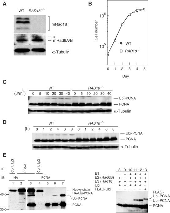
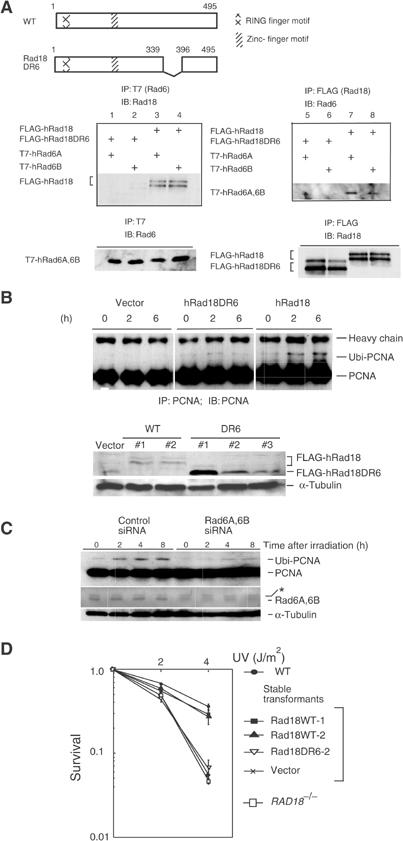
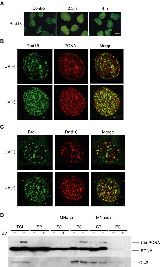
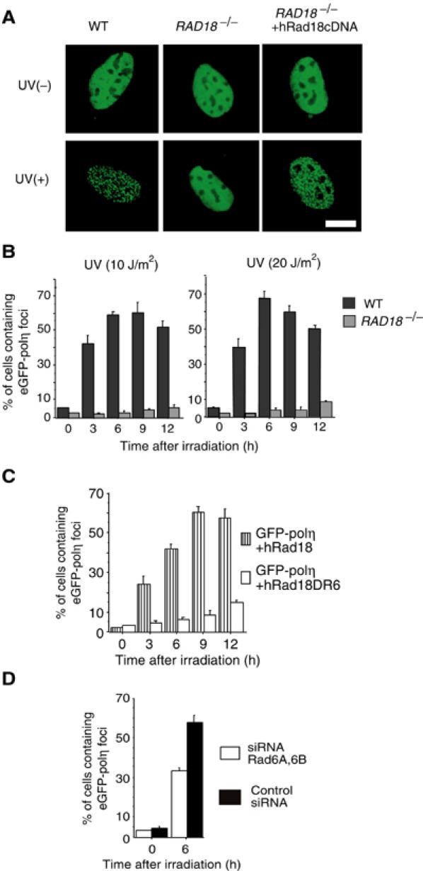
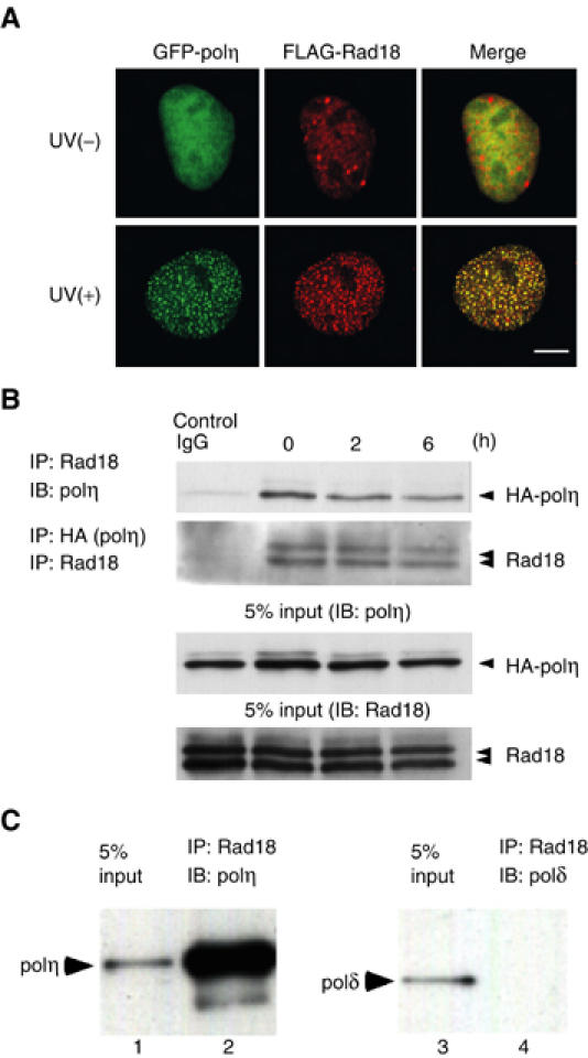
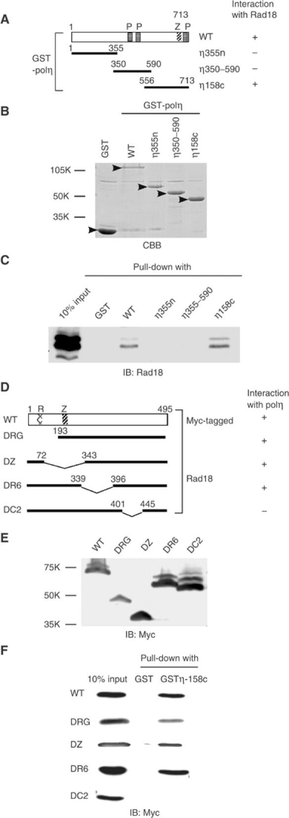
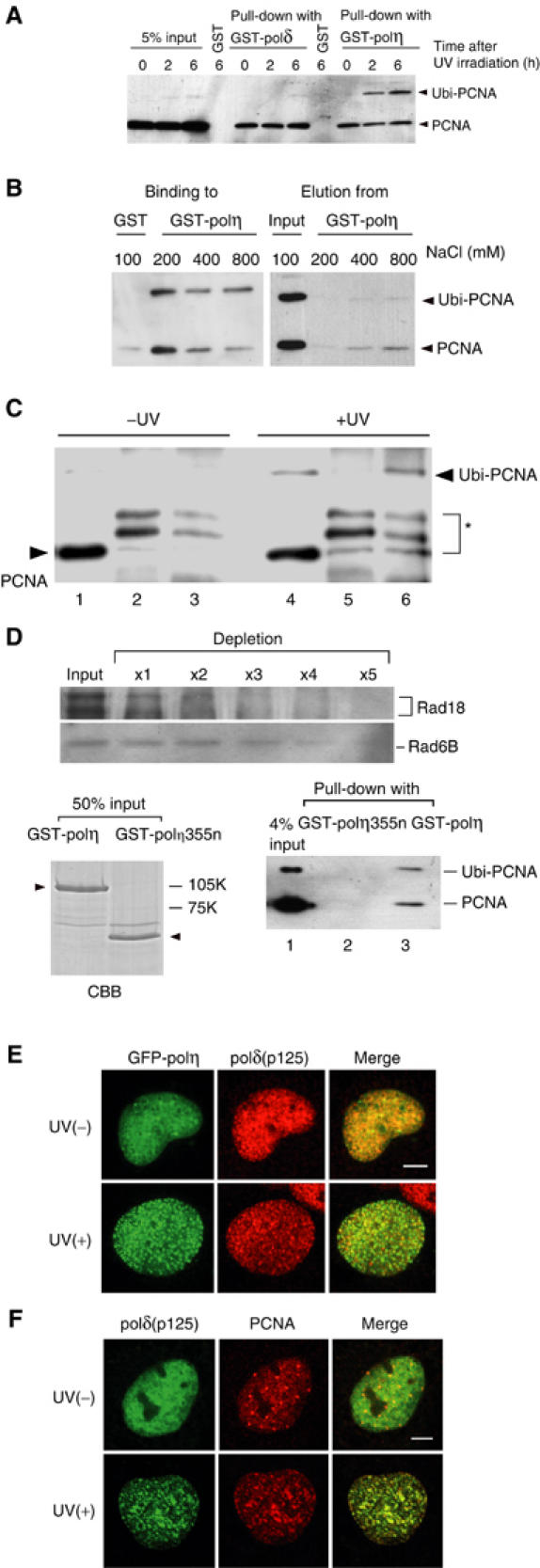
The DNA replication machinery stalls at damaged sites on templates, but normally restarts by switching to a specialized DNA polymerase(s) that carries out translesion DNA synthesis (TLS). In human cells, DNA polymerase eta (poleta) accumulates at stalling sites as nuclear foci, and is involved in ultraviolet (UV)-induced TLS. Here we show that poleta does not form nuclear foci in RAD18(-/-) cells after UV irradiation. Both Rad18 and Rad6 are required for poleta focus formation. In wild-type cells, UV irradiation induces relocalization of Rad18 in the nucleus, thereby stimulating colocalization with proliferating cell nuclear antigen (PCNA), and Rad18/Rad6-dependent PCNA monoubiquitination. Purified Rad18 and Rad6B monoubiquitinate PCNA in vitro. Rad18 associates with poleta constitutively through domains on their C-terminal regions, and this complex accumulates at the foci after UV irradiation. Furthermore, poleta interacts preferentially with monoubiquitinated PCNA, but poldelta does not. These results suggest that Rad18 is crucial for recruitment of poleta to the damaged site through protein-protein interaction and PCNA monoubiquitination.


Figures

Figure 1
Figure 1
Rad18 dependent monoubiquitination of PCNA by Rad18 and Rad6A/B in vivo and in vitro. (A) Western blot of Rad18 and Rad6A/B in RAD18−/− cells. α-Tubulin was included as a control. An asterisk shows nonspecific bands. (B) Growth curves of RAD18−/− cells. (C, D) Monoubiquitination of PCNA as determined by Western blot. Cells were harvested 5 h later following various doses of UV irradiation (C). In (D), cells were irradiated at 30 J/m2 and harvested at the indicated times. (E) In vivo (left, lanes 1–6) and in vitro (right, lanes 7–13) monoubiquitination of PCNA. GM637 cells were transfected with HA-ubiquitin (lanes 1–4) and irradiated with UV (13 J/m2, 6 h). Lysates were immunoprecipitated and blotted as indicated. In lanes 5 and 6, GM637 cells without transfection were irradiated at 0 and 13 J/m2 (6 h), respectively. Lane 7 represents an in vitro ubiquitination product. In lane 12, two-fold amounts of E2 and E3 were included in the reaction.
Figure 2
Figure 2
Requirement of Rad6A/B for monoubiquitination of PCNA in UV-irradiated cells. (A) Interaction of WT and mutant hRad18 with hRad6A/B. Full-length and mutant Rad18 proteins are schematically shown on the top panel. Plasmids were transfected into COS-7 cells with different combinations indicated on the left of the middle panels, and immunoprecipitation was performed. Similar levels of expression of hRad18 and hRad6A/B proteins in the transformed cells were confirmed in the lower panel. (B) Restoration of PCNA monoubiquitination in RAD18−/− cells by expression of WT hRad18 but not of mutant hRad18. Cells were incubated for 6 h following UV irradiation at 20 J/m2. Cell lysates were immunoprecipitated and blotted with an anti-PCNA antibody (upper panel). Expression of FLAG-hRad18 or FLAG-Rad18DR6 was confirmed in individual clones of stable transformants of RAD18−/− mouse fibroblasts by Western blot with an anti-Rad18 rabbit antibody (lower panel). α-Tubulin was indicated as a volume control. (C) Inhibition of PCNA monoubiquitination by siRNA for Rad6A/B. WI38VA13 cells were transfected with Rad6A and Rad6B siRNA, incubated for 4 days, and then irradiated with 10 J/m2 of UV light. At the indicated times, protein levels of monoubiquitinated PCNA were determined by Western blot. An asterisk shows a nonspecific band that remained constant following the siRNA treatment. (D) Restoration of UV sensitivity of RAD18−/− mouse cells by introduction of human Rad18 as determined by a colony-forming assay. Two independent clones of stable transformants (WT#1 and WT#2 in (B)) were tested.
Figure 3
Figure 3
Colocalization of Rad18 with PCNA on chromatin following UV irradiation. (A) Dispersion and relocalization of Rad18. GM637 cells irradiated at 15 J/m2 were fixed with formaldehyde and stained for Rad18. Bar=20 μm. (B) UV-induced colocalization of Rad18 with PCNA. GM637 cells irradiated at 15 J/m2 were fixed with methanol 4 h after UV irradiation and processed for double staining for Rad18 (green) and PCNA (red). Bar=10 μm. (C) Accumulation of Rad18 at the replication stalling sites. UV-irradiated (15 J/m2) GM637 cells were labeled for 2 h with BrdU, fixed with methanol, and processed for double staining for Rad18 (red) and BrdU (green). Bar=10 μm. (D) Binding of monoubiquitinated PCNA to chromatin. Chromatin fractions were isolated from UV-irradiated (15 J/m2, 6 h) or nonirradiated HeLa cells, and then treated with micrococcal nuclease (MNase). The distributions of PCNA in the total cell lysate (TCL), soluble fraction (S2), solubilized nuclear fraction (S3), and chromatin-enriched fraction (P3) are shown. Orc2 is shown as a chromatin fraction marker.
Figure 4
Figure 4
Rad18- and Rad6-dependent formation of polη foci. (A) Focus formation of eGFP-polη following UV irradiation in WT cells but not in RAD18−/− cells. Cells were irradiated at 15 J/m2. After 6 h, the distribution of eGFP-polη was examined after fixation. Defective focus formation of polη was recovered by concomitant expression of Rad18. Bar=10 μm. (B) Time course of eGFP-polη focus formation in UV-irradiated cells. RAD18−/− mouse cells and WT cells were transfected with eGFP-polη. After 20 h, cells were irradiated with UV at the indicated doses. (C) Restoration of eGFP-polη focus formation in UV-irradiated (20 J/m2) RAD18−/− cells by expression of WT hRad18 but not of mutant hRad18 lacking the Rad6-binding domain. (D) Inhibition of polη focus formation by siRNA for Rad6. WI38VA13 cells were transfected with Rad6A and Rad6B siRNA, cultured for 3 days, and then transfected again with an eGFP-polη plasmid. After 20 h, cells were irradiated with UV (10 J/m2), and 6 h later cells containing eGFP-polη foci were counted.
Figure 5
Figure 5
Direct interaction of polη with Rad18. (A) UV-induced colocalization of Rad18 with eGFP-polη in GM637 cells. Cells transfected with eGFP-polη and FLAG-Rad18 plasmids were irradiated at 15 J/m2 and incubated for 6 h. After fixation, cells were stained for Rad18 with an antibody against FLAG. Bar=10 μm. (B) Interaction of Rad18 with polη. HA-polη was transiently expressed in GM637 cells. Immunoprecipitation was performed at various times after UV irradiation (12.5 J/m2). As a control, UV-irradiated cell lysates (6 h) were immunoprecipitated with control IgG. (C) Direct binding of Rad18 with polη. Recombinant Rad18 and polη were purified from insect cells. After incubation of the mixture, Rad18 was immunoprecipitated and polη bound to Rad18 was detected by Western blot. Polδ was used as a control.
Figure 6
Figure 6
Determination of binding sites. (A) Structural domains of GST-polη fusion proteins. P: putative PCNA-binding domain; Z: zinc-finger domain. (B) Purification of GST- polη fusion proteins by glutathione beads. Proteins bound to the beads were stained with Coomassie brilliant blue (CBB, arrowheads). (C) Pull-down assay. GM637 cell lysates were pulled down with GST-polη fusion proteins bound to glutathione beads. Interaction with Rad18 was analyzed by Western blot. (D) Structural domains of Myc-tagged Rad18 proteins. R: RING finger domain; Z: zinc-finger domain. (E) Deletion mutant proteins were overexpressed in COS-7 cells and their expression was confirmed by Western blot. (F) COS-7 cell lysates containing Myc-tagged mutant Rad18 proteins were pulled down with GST-polη158c bound to glutathione beads. Association of WT and mutant Rad18 proteins with polη was analyzed by Western blot using an anti-Myc antibody.
Figure 7
Figure 7
Preferential binding of polη to monoubiquitinated PCNA. (A) Binding of polη to ubiquitinated PCNA. HeLa cells were irradiated with UV at 20 J/m2. PCNA in the cell lysates was pulled down with either GST-polη beads or polδ beads, and analyzed by Western blot using an anti-PCNA antibody. (B) Effects of different salt concentrations on the binding of PCNA to GST-polη (left) and on PCNA elution from GST-polη (right). PCNA pulled down was washed with buffer containing various concentrations of NaCl. PCNA in bound or eluted fractions was analyzed as in (A). (C) Preferential binding of polη to monoubiquitinated PCNA in living cells. GM637 cells were transfected with an HA-polη plasmid. After 2 days, these cells were irradiated with 20 J/m2 of UV (lanes 4–6), or remained untreated (lanes 1–3), and incubated for 5 h. After immunoprecipitation with an anti-polη antibody (lanes 3 and 6) or control IgG (lanes 2 and 5), binding PCNA was detected by Western blot with an anti-PCNA antibody. Lanes 1 and 4 represent 5% samples of the whole-cell lysate. An asterisk shows nonspecific bands. (D) Direct binding of polη to monoubiquitinated PCNA. Rad18 was removed from the in vitro PCNA ubiquitination reaction mixture by immunoprecipitation with an anti-Rad18 antibody (upper panel). Note that Rad6B was also depleted, probably due to direct interaction with Rad18 (upper). Remaining PCNA and monoubiquitinated PCNA were pulled down with purified GST-polη bound to glutathione beads (lower, right). GST-polη355n was used as a control. Purity of the polymerase samples is shown on the left panel (arrowheads) by the Coomassie brilliant blue (CBB) staining. (E) Colocalization of eGFP-polη with polδ in UV-irradiated (10 J/m2, 5 h) GM637 cells. Bar=5 μm. (F) Colocalization of polδ with PCNA in UV-irradiated (10 J/m2, 5 h) WI38VA13 cells. Bar=5 μm.

References

    1. Bailly V, Lamb J, Sung P, Prakash S, Prakash L (1994) Specific complex formation between yeast RAD6 and RAD18 proteins: a potential mechanism for targeting RAD6 ubiquitin-conjugating activity to DNA damage sites. Genes Dev 8: 811–820 - PubMed
    1. Bailly V, Lauder S, Prakash S, Prakash L (1997a) Yeast DNA repair proteins Rad6 and Rad18 form a heterodimer that has ubiquitin conjugating, DNA binding, and ATP hydrolytic activities. J Biol Chem 272: 23360–23365 - PubMed
    1. Bailly V, Prakash S, Prakash L (1997b) Domains required for dimerization of yeast Rad6 ubiquitin-conjugating enzyme and Rad18 DNA binding protein. Mol Cell Biol 17: 4536–4543 - PMC - PubMed
    1. Broomfield S, Hryciw T, Xiao W (2001) DNA postreplication repair and mutagenesis in Saccharomyces cerevisiae. Mutat Res 486: 167–184 - PubMed
    1. Broughton BC, Cordonnier A, Kleijer WJ, Jaspers NGJ, Fawcett H, Raams A, Garritsen VH, Stary A, Avril MF, Boudsocq F, Masutani C, Hanaoka F, Fuchs RP, Sarasin A, Lehmann AR (2002) Molecular analysis of mutation in DNA polymerase η in xeroderma pigmentosum-variant patients. Proc Natl Acad Sci USA 99: 815–820 - PMC - PubMed

Publication types

MeSH terms