. 2004 Oct 1;23(19):3897-906.
doi: 10.1038/sj.emboj.7600395. Epub 2004 Sep 9.

Requirement of IS911 replication before integration defines a new bacterial transposition pathway

Affiliations

Requirement of IS911 replication before integration defines a new bacterial transposition pathway

G Duval-Valentin et al. EMBO J. .

Abstract

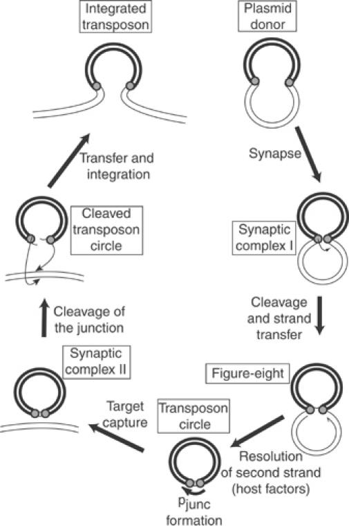
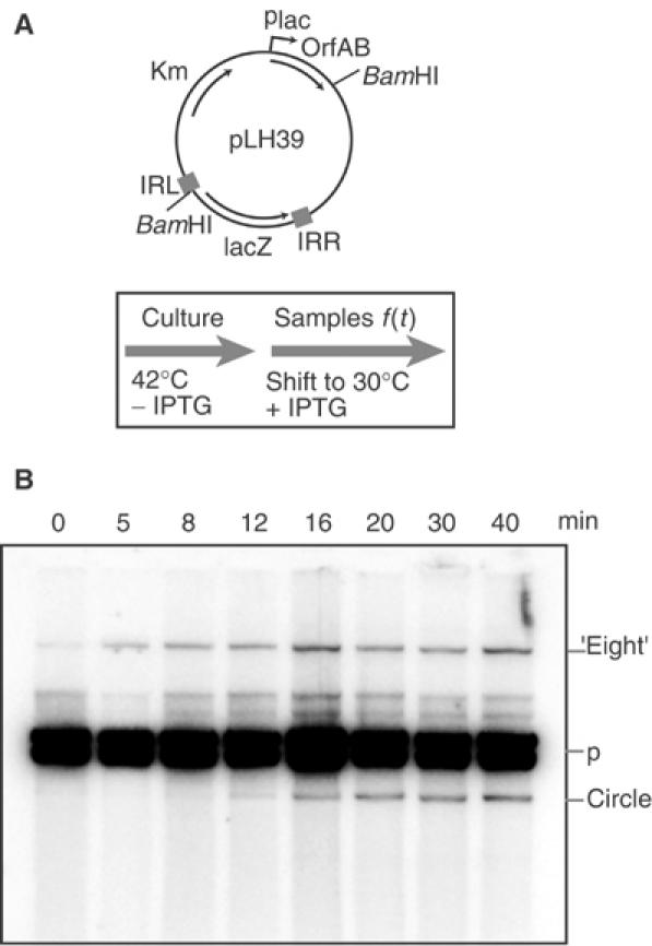
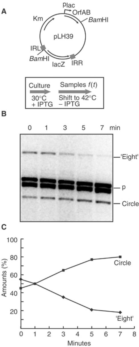
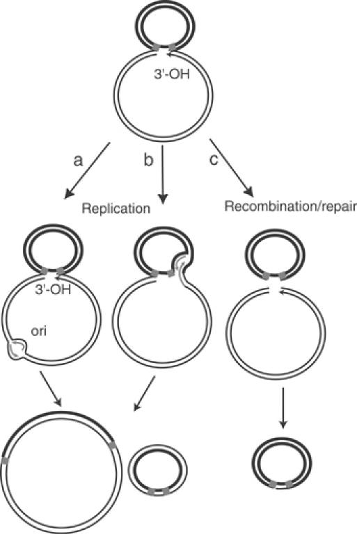
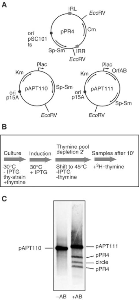
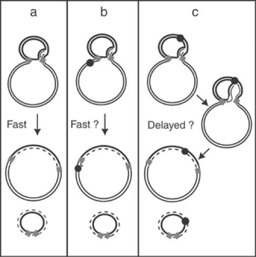
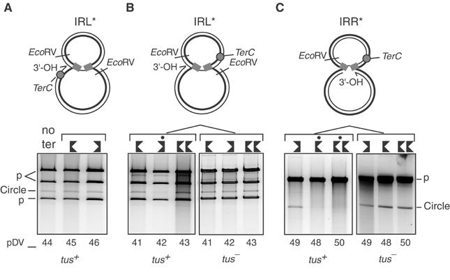
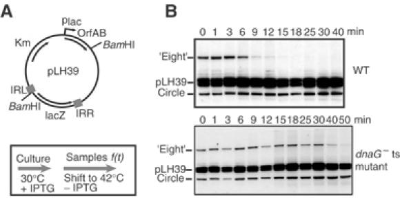
Movement of transposable elements is often accompanied by replication to ensure their proliferation. Replication is associated with both major classes of transposition mechanisms: cut-and-paste and cointegrate formation (paste-and-copy). Cut-and-paste transposition is often activated by replication of the transposon, while in cointegrate formation replication completes integration. We describe a novel transposition mechanism used by insertion sequence IS911, which we call copy-and-paste. IS911 transposes using a circular intermediate (circle), which then integrates into a target. We demonstrate that this is derived from a branched intermediate (figure-eight) in which both ends are joined by a single-strand bridge after a first-strand transfer. In vivo labelling experiments show that the process of circle formation is replicative. The results indicate that the replication pathway not only produces circles from figure-eight but also regenerates the transposon donor plasmid. To confirm the replicative mechanism, we have also used the Escherichia coli terminators (terC) which, when bound by the Tus protein, inhibit replication forks in a polarised manner. Finally, we demonstrate that the primase DnaG is essential, implicating a host-specific replication pathway.


Figures

Figure 1
Figure 1
IS911 transposition pathway. The transposon (heavy line), plasmid backbone (lighter line), and transposon termini (grey circles) are indicated. The transposon ends are brought together in a synapse (synaptic complex I). A first single-strand cleavage occurs initially at one end of the IS, catalysed by the low level of Tpase expressed from a weak promoter, pIRL, present in IRL. The free 3′-OH generated is then directed to attack the opposite end on the same DNA strand (strand transfer) to generate a figure-eight form in which both ends are joined by a single-strand bridge, leaving an unjoined DNA strand with a free 3′-OH (half arrow) on the vector plasmid. The figure-eight is converted into a transposon circle where the formation of a strong promoter, pjunc, induces high-level synthesis of transposon proteins (Ton-Hoang et al, 1997; Duval-Valentin et al, 2001). A second Tpase-mediated synaptic complex (synaptic complex II) is formed upon target capture. The Tpase then undergoes the final steps of cleavage and strand transfer into the target DNA (integration). Insertion results in the disassembly of the strong promoter and returns to a low level of transposition activity.
Figure 2
Figure 2
In vivo kinetics of figure-eight and circle formation. (A) Plasmid structure and experimental regime. Plasmid pLH39 is a p15A-based plasmid carrying an artificial IS911-derived transposon and an independent OrfAB gene under control of placuv5. The position of BamHI sites to separate different molecules is shown. The culture was grown at 42°C (OrfAB activity is temperature sensitive) to ensure that there was no residual Tpase activity. At t=0 (OD600=0.6), the population was transferred into LB medium prewarmed at 30°C and production of Tpase was induced with IPTG. Samples were withdrawn at different times, growth was stopped by addition of an sodium azide/ice mixture, and cleared lysates were then made. (B) Separation of plasmid species. Agarose gel of BamHI-digested DNA samples removed at times 0–40 min (shown above the lanes). The separated species were visualised by hybridisation directly in the dried gel with a transposon-specific oligonucleotide probe. The position of the χ forms generated by BamHI digestion of the figure-eight molecules is shown (‘eight'), together with the two bands obtained from the donor plasmid (p) and the BamHI-linearised transposon circles. Note that some additional minor bands are due to incomplete digestion of plasmid molecules.
Figure 3
Figure 3
In vivo kinetics of figure-eights to circles conversion. (A) Plasmid structure and experimental regime. The plasmid used was the same as described in Figure 2 (A). The culture was grown at 30°C in the presence of IPTG to induce Tpase synthesis and accumulate figure-eight. At t=0, the culture was transferred into fresh LB medium prewarmed at 42°C without IPTG to inactivate Tpase and stop the induction. Samples were withdrawn at different times and treated as described in Figure 2. (B) Separation of plasmid species. Agarose gel showing details of samples taken between 0 and 7 min. The separated species were visualised by hybridisation as in Figure 2. The symbols are as described in Figure 2. (C) Quantitation of gel shown in (B). The values shown were calculated as follows: the intensities of figure-eight and circle bands were measured and the sum was used to define a value of 100%. The level of parental plasmid was used to normalise sampling variations from well to well.
Figure 4
Figure 4
Model of three possible pathways for resolving figure-eight molecules to circles. The figure-eight molecule carrying a 3′-OH is shown above. The two left-hand drawings show circle formation driven by replication from the plasmid origin of replication (left) or from the Tpase-generated 3′-OH (centre). In both cases, replication would generate a double-stranded transposon circle and regenerate the original parental plasmid. Conversion using a recombination/repair pathway is shown on the right. Host-mediated repair would be expected to lead to either destruction or recircularisation of the donor plasmid backbone.
Figure 5
Figure 5
3H-thymidine incorporation in vivo during circle formation. (A) Plasmid structure. The temperature-sensitive plasmid replication mutant, pPR4, carrying the IS911-based transposon with a chloramphenicol resistance gene, Cm, located between correctly oriented IS911 ends (IRL and IRR), is shown together with both the p15A-derived Tpase vector plasmid, pAPT111, carrying the orfAB gene, and its isogenic parent, pAPT110, lacking this gene. (B) Experimental regime. A culture of a thyA strain carrying a temperature-sensitive pSC101 derivative as a transposon vector (pPR4) and either pAPT110 or pAPT111 was grown at 30°C in minimal medium complemented with thymine, and OrfAB synthesis was induced with IPTG. After growth to OD600=0.6, the culture was transferred into fresh medium without thymine or IPTG at 45°C in order to rapidly inactivate OrfAB production and replication of the transposon-carrying plasmid. After 2 min of incubation to deplete the internal pool of thymine, labelling with 3H-thymidine was performed for 10 min, alkaline lysates were prepared, plasmid DNA was digested with EcoRV and analysed by agarose gel electrophoresis. (C) Agarose gel electrophoresis. The species were visualised directly by radio-luminescence as described in Materials and methods. Left lane: control experiment in the absence of OrfAB. Only the p15A-based vector plasmid pAPT110 is labelled, confirming that the temperature-sensitive pPR4 is not replicated at the nonpermissive temperature and that no circles were detected. Right lane: both the OrfAB-carrying p15A-based vector plasmid and the temperature-sensitive pPR4 plasmid with the transposon are labelled.
Figure 6
Figure 6
Scheme showing experiments to confirm the replicative pathway of figure-eight resolution in the presence of different locations of terC sites. ter sites and the Tus protein inhibit replication fork movement in a polar manner. terC sequences were therefore introduced either outside (scheme b) or inside (scheme c) an IS911-derived transposon in both orientations. The kinetics of circle formation would be expected to be affected differentially, depending on the orientation of ter. The predicted effects on the resolution rate are proposed. The newly replicated strand is shown as a dotted line.
Figure 7
Figure 7
terC affects figure-eight resolution in a polar manner. IR donor mutants were used to obtain unique figure-eight populations and to orient the potential replication fork (AC, top). The 1% agarose gels were stained with Sybr green. IRL* or IRR* donor mutants (A–C, bottom) were tested separately with different positions (shown as grey circles, top) and orientations of terC sites (shown by oriented black symbols, bottom; Table I). These were placed outside the transposon (pDV45 and 46 with IRL*; A) or inside the transposon with IRL* (pDV41, 42, 43; B) or with IRR* (pDV48, 49, 50; C). pDV43 and 50 harbour two terC sites in tandem. The right-hand panels (B, C) show the results obtained in a tus strain. The major upper band marked p in (A, B) represents linearised pAPT111. The lower two bands marked p (A, B) represent two fragments generated from the transposon-carrying plasmids. Introduction of IRR* (C) resulting in the elimination of one of the two EcoRV sites from the parental plasmid pRP4 (Figure 5). These plasmids are therefore linearised by EcoRV and migrate at the same position as the linearised Tpase donor plasmid. The band corresponding to plasmid circles is also indicated. Additional minor bands migrate at positions consistent with partially digested products. Lanes in which transposon circle formation was delayed are indicated (*).
Figure 8
Figure 8
In vivo kinetics of figure-eight to circle conversion in a dnaGts strain. (A) Plasmid structure and experimental regime. After production of figure-eight molecules at 30°C (Tpase OrfAB and DnaG active (A)), the conversion of figure-eights to circles of a wild-type strain was compared with the dnaGts strain at 42°C. Samples were withdrawn at different times and treated as described in Figure 2. (B) Separation of plasmid species by agarose gel electrophoresis. The top panel shows results obtained with the wild-type strain, while the lower panel presents the results obtained with the dnaG mutant. Numbers above the gel indicate the time in minutes after the temperature shift.

References

    1. Birnboim HC, Doly J (1979) A rapid alkaline extraction procedure for screening recombinant plasmid DNA. Nucleic Acids Res 7: 1513–1523 - PMC - PubMed
    1. Bussiere DE, Bastia D (1999) Termination of DNA replication of bacterial and plasmid chromosomes. Mol Microbiol 31: 1611–1618 - PubMed
    1. Chaconas G, Harshey RM (2002) Transposition of phage Mu DNA. In: Mobile DNA II, Craig N, Craigie R, Gellert M, Lambowitz A (eds), Washington, DC: ASM Press
    1. Chandler M, Mahillon J (2002) Insertion sequences revisited. In Mobile DNA II, Craig N, Craigie R, Gellert M, Lambowitz A (eds), pp 305–366. Washington, DC: ASM Press
    1. Churchward G, Belin D, Nagamine Y (1984) A pSC101-derived plasmid which shows no sequence homology to other commonly used cloning vectors. Gene 31: 165–171 - PubMed

Publication types