. 2005 Jan 1;385(Pt 1):289-99.
doi: 10.1042/BJ20040371.

Ribosomal protein L7a binds RNA through two distinct RNA-binding domains

Affiliations

Ribosomal protein L7a binds RNA through two distinct RNA-binding domains

Giulia Russo et al. Biochem J. .

Abstract

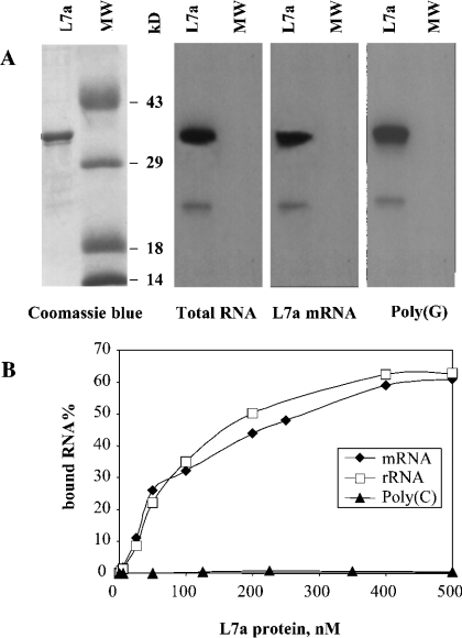
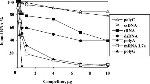
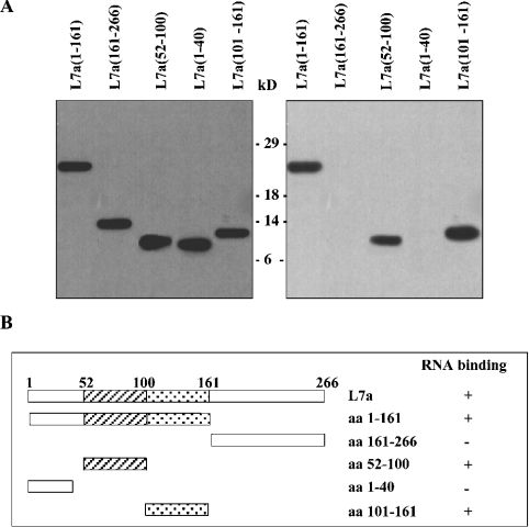
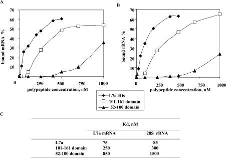
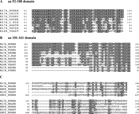
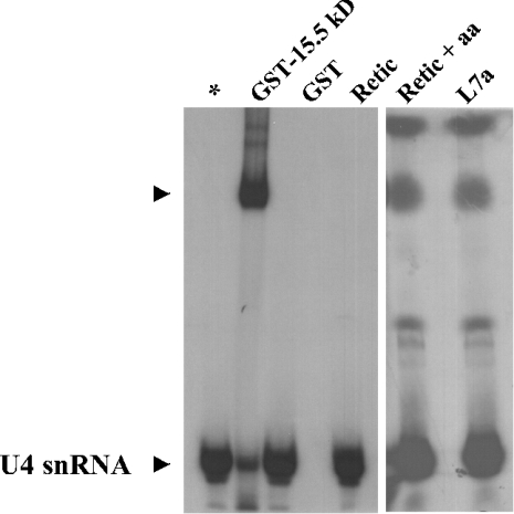
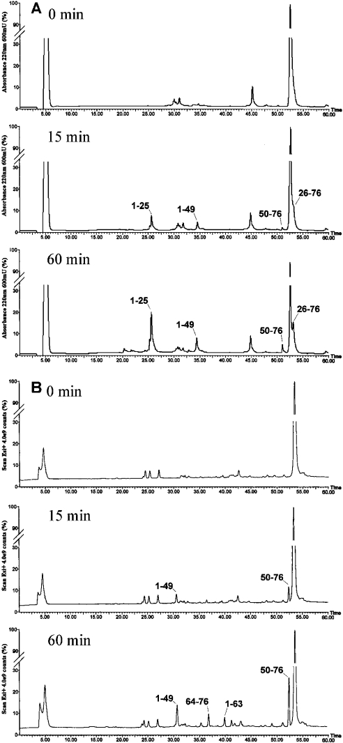
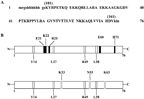
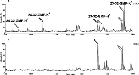
The human ribosomal protein L7a is a component of the major ribosomal subunit. We previously identified three nuclear-localization-competent domains within L7a, and demonstrated that the domain defined by aa (amino acids) 52-100 is necessary, although not sufficient, to target the L7a protein to the nucleoli. We now demonstrate that L7a interacts in vitro with a presumably G-rich RNA structure, which has yet to be defined. We also demonstrate that the L7a protein contains two RNA-binding domains: one encompassing aa 52-100 (RNAB1) and the other encompassing aa 101-161 (RNAB2). RNAB1 does not contain any known nucleic-acid-binding motif, and may thus represent a new class of such motifs. On the other hand, a specific region of RNAB2 is highly conserved in several other protein components of the ribonucleoprotein complex. We have investigated the topology of the L7a-RNA complex using a recombinant form of the protein domain that encompasses residues 101-161 and a 30mer poly(G) oligonucleotide. Limited proteolysis and cross-linking experiments, and mass spectral analyses of the recombinant protein domain and its complex with poly(G) revealed the RNA-binding region.


Figures

Figure 1
Figure 1. Analysis of RNA-binding ability of human r-protein L7a
Purified recombinant L7a–His protein (L7a) and molecular mass marker proteins (MW) were loaded on to an SDS gel. Upon electrophoresis, gel samples were stained with Coomassie Brilliant Blue (A, left-hand panel), or transferred on to a nitrocellulose membrane, probed with the indicated radiolabelled RNAs and subjected to autoradiography (A, right-hand panels; for an explanation for the lanes marked MW see the text). (B) Plot of binding activity of L7a–His to radiolabelled RNAs. For details of nucleic acid labelling and filter-binding assay, see the Experimental section.
Figure 2
Figure 2. RNA-binding specificity of human r-protein L7a
Filter-binding assay of competition of the binding of L7a–His to radiolabelled L7a mRNA by increasing amounts of unlabelled nucleic acids. The binding reaction contained 10 fmol of labelled L7a mRNA, to which a competitor nucleic acid was added before the addition of 500 nM L7a–His. The assay was performed as described in the Experimental section. The bound RNA values were corrected for the radioactivity retained on the filter when no L7a protein was added to the labelled L7a mRNA. The RNA-binding activity of L7a protein to radiolabelled L7a mRNA in the absence of a competitor was considered as 100%.
Figure 3
Figure 3. Definition of RNA-binding domains of the human r-protein L7a
(A) Purified recombinant L7a-His deletion mutants were subjected to electrophoresis on an SDS/PAGE gel and then transferred on to a nitrocellulose membrane. The filter was probed with a commercially available antiserum against the His-tag (Santa Cruz Biotechnology) (left-hand panel). The RNA-binding activity of all deletion mutants was tested by Northwestern experiments using radiolabelled L7a mRNA (right-hand panel). (B) Summary of the RNA-binding ability of L7a deletion mutants. Striped boxes indicate the domain required for nucleolar targeting of the protein, and dotted regions indicate the putative RNA-binding motif [10].
Figure 4
Figure 4. RNA-binding activity of L7a–His, and L7a-derived polypeptides with L7a mRNA (A) and 28 S RNA (B)
The filter-binding assays were carried out as described in the Experimental section. The resulting plots are the average of four independent experiments and constitute the basis for the calculation of the Kd values reported in (C).
Figure 5
Figure 5. Evolutionary conservation of the RNA-binding domains of r-protein L7a
(A) Alignment of the RNAB1 (aa 52–100) domain of the human L7a r-protein (RL7A_HUMAN; UniProt database accession number P11518) with the orthologue protein from chicken (RL7A_CHICK; P32429), Fugu rubripes (RL7A_FUGRU; O57592), Drosophila melanogaster (RL7A_DROME; P46223), Caenorhabditis elegans (RL7A_C. ELE; Q8T875), Arabidopsis thaliana (RL7A_ARATH; P49692), Oryza sativa (RL7A_ORYSA; P35685) and Saccharomyces cerevisiae (RL8B_YEAST; P29453). (B) Alignment of the RNAB2 (aa 101–161) domain of the human r-protein L7a with the eukaryotic orthologues shown in (A), and with the Archea H. marismortui (RL7A_HALMA; P12743) and P. abyssi (RL7A_PYRHO; O59165) orthologues. (C) Sequence alignment of the extended RNAB2 domain of human r-protein L7a with: Archea L7ae protein, human 15.5 kDa protein (NHPX_HUMAN; P55769), yeast L30 (RL30_YEAST; P14120), human SBP2 (SBP2_HUMAN; Q96T21). Identical residues are in black and conserved residues are grey boxes. The conserved residues are grouped DE, KRH, LIVAMPYFW and TSQCN.
Figure 6
Figure 6. EMSA analysis of binding of proteins 15.5 kDa and L7a to U4 snRNA 5′-stem-loop oligonucleotide
The recombinant GST–15.5 kDa fusion protein, or L7a protein produced by in vitro translation were incubated with the 5′-end labelled U4 snRNA oligonucleotide, and the protein–RNA complex was resolved by PAGE on a 6% native polyacrylamide gel. As a control, the EMSA was performed with the probe alone (*), with recombinant GST protein (GST), with unprogrammed rabbit reticulocyte lysate (Retic), and with unprogrammed rabbit reticulocyte lysate plus amino acids (Retic+aa).
Figure 7
Figure 7. Limited proteolysis of native L7a(101–161) (A), and the L7a(101–161)-poly(G) complex (B) with trypsin
HPLC analysis of the native L7a(101–161) and of the L7a(101–161)–poly(G) complex after 0 min, 15 min and 60 min of incubation with trypsin. The indicated fractions were manually collected and the eluted peptides were identified by ESMS in the case of the isolated protein, and directly by the LCMS procedure in the case of the complex.
Figure 8
Figure 8. Location of proteolytic sites in the native L7a(101–161) protein and in the L7a(101–161)–poly(G) complex
(A) Sequence of the L7a(101–161) recombinant polypeptide. Capital letters indicate the amino acid residues of L7a; the corresponding position in the protein is indicated in parentheses. Lower case letters indicate the amino acid residues added by the cloning strategy. (B) Schematic representation of the results obtained from limited proteolysis experiments on L7a(101–161) in the absence (upper panel) and in the presence of poly(G) (lower panel). Preferential proteolytic sites occurring only in the native protein are shown in black; those recognized by proteases in the complex are shown in grey, and those identified in all conditions are shown in white.
Figure 9
Figure 9. Cross-linking experiment on the L7a(101–161)–poly(G) complex
Partial MALDI-MS spectrum of the cross-linked product (A) and of the control isolated protein (B) digested with trypsin and T1 ribonuclease. The attribution of the cross-linked peptides is shown.

References

    1. Hernandez-Verdun D., Roussel P., Gebrane-Younes J. Emerging concepts of nucleolar assembly. J. Cell Sci. 2002;115:2265–2270. - PubMed
    1. Rudt F., Pieler T. Cytosolic import factor- and Ran-independent nuclear transport of ribosomal protein L5. Eur. J. Cell Biol. 2001;80:661–668. - PubMed
    1. Russo G., Ricciardelli G., Pietropaolo C. Different domains cooperate to target the human ribosomal L7a protein to the nucleus and to the nucleoli. J. Biol. Chem. 1997;272:5229–5235. - PubMed
    1. Damelin M., Silver P. A., Corbett A. H. Nuclear protein transport. Methods Enzymol. 2002;351:587–607. - PubMed
    1. Jakel S., Mingot J. M., Schwarzmaier P., Hartmann E., Gorlich D. Importins fulfil a dual function as nuclear import receptors and cytoplasmic chaperones for exposed basic domains. EMBO J. 2002;21:377–386. - PMC - PubMed

Publication types

MeSH terms

LinkOut - more resources