Comparative Study
. 2004 Oct;5(10):1211-7.
doi: 10.1631/jzus.2004.1211.

Ultrastructure of spermatozoa of Bullacta exarata (philippi) and its significance on reproductive evolution and physio-ecological adaptation

Affiliations
Comparative Study

Ultrastructure of spermatozoa of Bullacta exarata (philippi) and its significance on reproductive evolution and physio-ecological adaptation

Xue-ping Ying et al. J Zhejiang Univ Sci. 2004 Oct.

Abstract

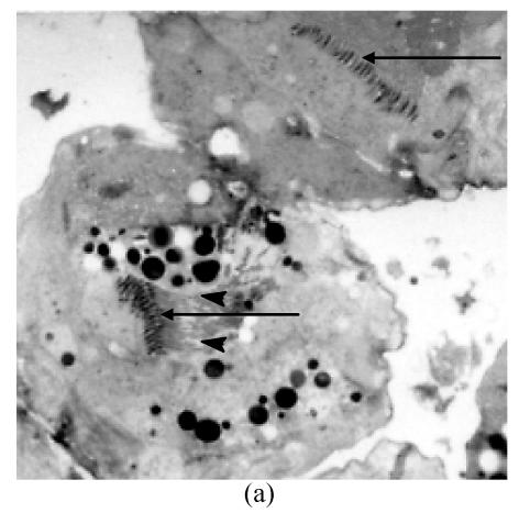
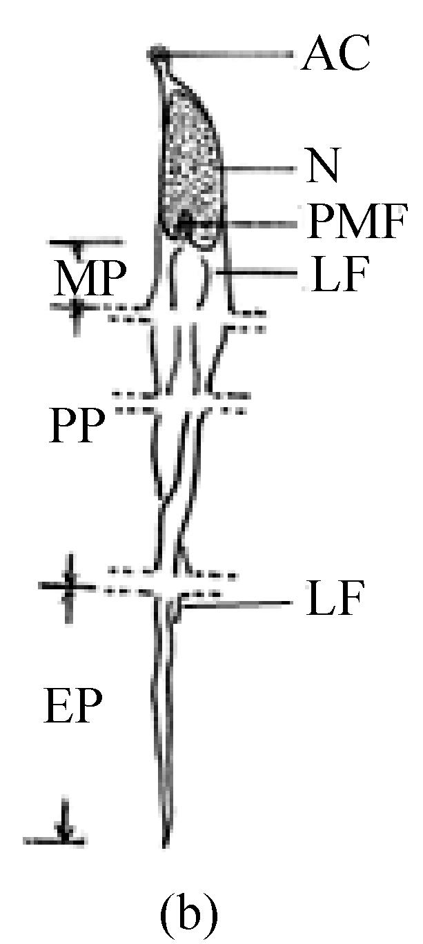
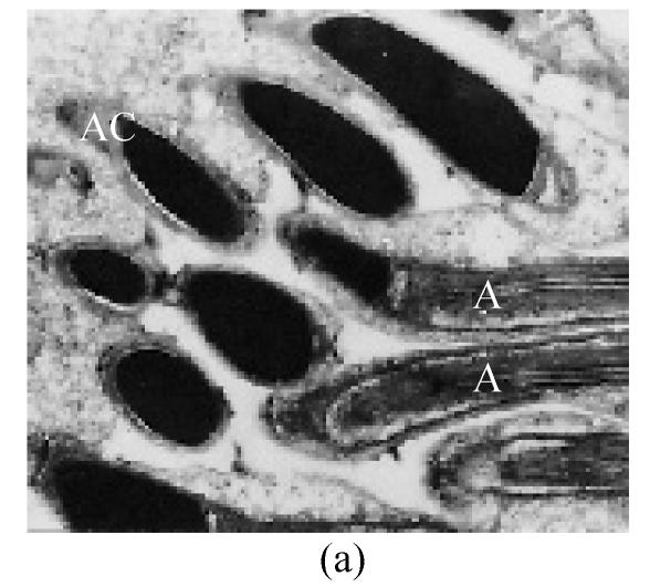
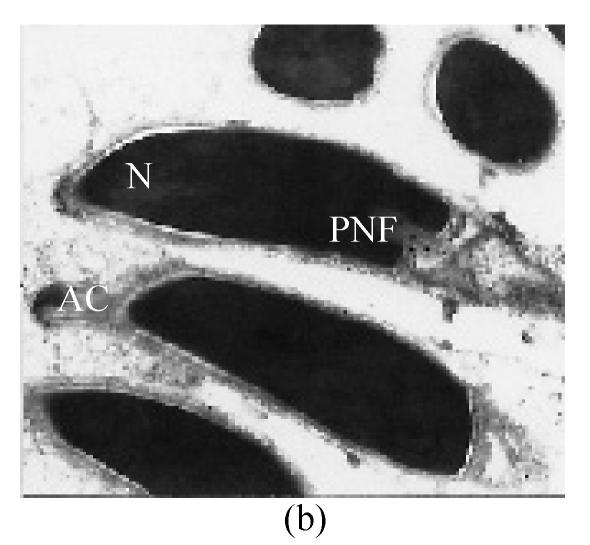
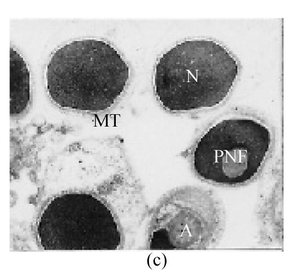
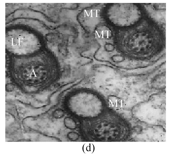
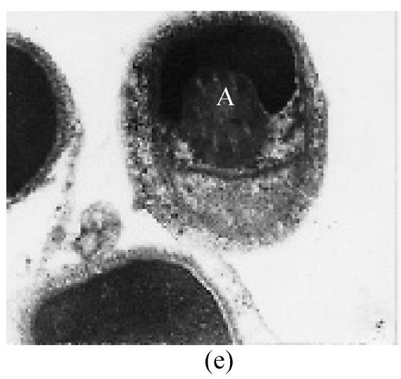
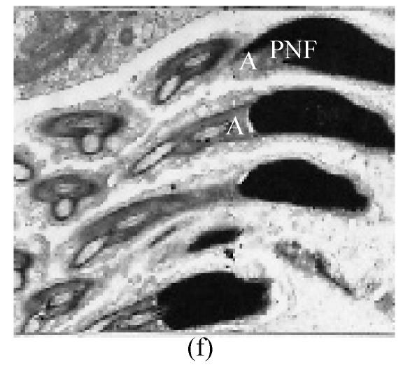
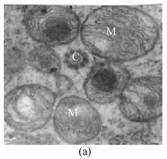
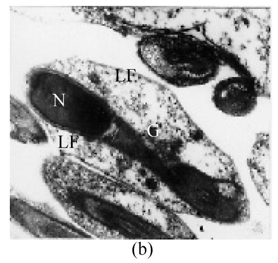
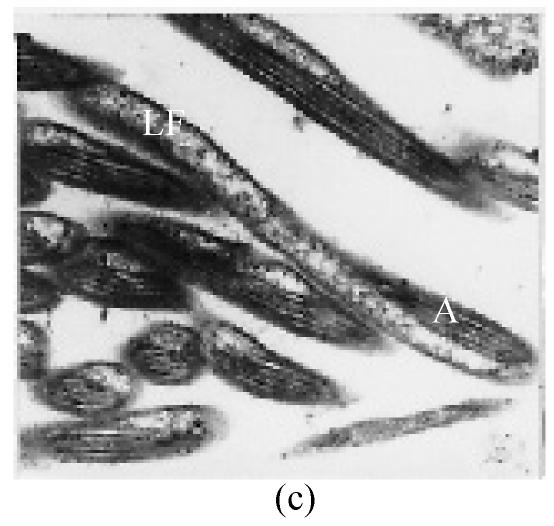
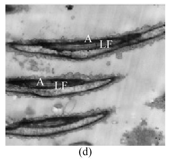
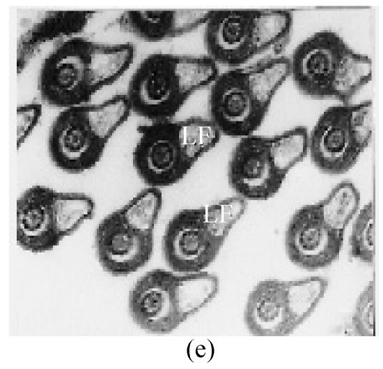
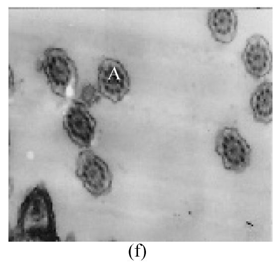
The morphology and ultrastructure of Bullacta exarata spermatozoa observed by light and transmission electron microscopy are presented in this paper. The spermatozoa is composed of head with a simple acrosomal complex and an elongated nucleus, and tail with a midpiece, principal piece and an end piece. The midpiece consists of a mitochondrial ring, and the principal piece is composed of axoneme and lateral fin. The structure of B. exarata spermatozoa differs significantly from that of other gastropods, especially in the lateral fin and the principal piece, which was described scarcely before. A comparison is made between B. exarata and other gastropods, and its significance on reproductive evolution and physio-ecological adaptation is preliminarily discussed.


Figures

Fig. 1
Fig. 1
Mature testis of B. exarata was semi-sectioned and stained with Toluidine Blue. The nuclei are clear, and arranged in parallel (a, arrows); the tails are indicated by arrow heads. Two parts of the spermatozoon are fully labeled in the diagram (b) AC, acrosome; EP, end piece; LF, lateral fin; MP, mid-piece; N, nucleus; PNF, posterior nuclear fossa; PP, principal piece
Fig. 1
Fig. 1
Mature testis of B. exarata was semi-sectioned and stained with Toluidine Blue. The nuclei are clear, and arranged in parallel (a, arrows); the tails are indicated by arrow heads. Two parts of the spermatozoon are fully labeled in the diagram (b) AC, acrosome; EP, end piece; LF, lateral fin; MP, mid-piece; N, nucleus; PNF, posterior nuclear fossa; PP, principal piece
Fig. 2
Fig. 2
Ultrastructure of B. exarata (Philippi) spermatozoa (a) Mature spermatozoa of Bullacta exarata (×8000); (b) Mature acrosomal complex, nuclear and posterior nuclear fossa (×15000); (c) Transverse section of nucleus surrounded by microtubules (×20000); (d) Transverse section of an imature sperm, showing microtubules, lateral fins and the “9+2” structure (×35000); (e) Transverse section of nucleas, also showing the axoneme (×35000); (f) Longitudinal section of spermatozoa, showing axoneme and posterior nuclear fossa (×8000) A: axoneme; AC: acrosome cap; LF: lateral fin; MT: microtubules; N: nucleus; PNF: posterior nuclear fossa
Fig. 2
Fig. 2
Ultrastructure of B. exarata (Philippi) spermatozoa (a) Mature spermatozoa of Bullacta exarata (×8000); (b) Mature acrosomal complex, nuclear and posterior nuclear fossa (×15000); (c) Transverse section of nucleus surrounded by microtubules (×20000); (d) Transverse section of an imature sperm, showing microtubules, lateral fins and the “9+2” structure (×35000); (e) Transverse section of nucleas, also showing the axoneme (×35000); (f) Longitudinal section of spermatozoa, showing axoneme and posterior nuclear fossa (×8000) A: axoneme; AC: acrosome cap; LF: lateral fin; MT: microtubules; N: nucleus; PNF: posterior nuclear fossa
Fig. 3
Fig. 3
Ultrastructure of midpiece, principal and end piece of B. exarata (Philippi) spermatozoa (a) showing the mitochondria around the centriole (×40000); (b) Showing the lateral fins in the two sides of midpiece (×17000); (c) Longitudinal section of principal piece, showing the axoneme and lateral fins twisting together (×15000); (d) Longitudinal section of the sperm tail, showing the axoneme and lateral fins (×12000); (e) Transverse section of the sperm tail, showing the structure of the lateral fins (×25000); (f) Transverse section of the end piece of the sperm (×25000) A: axoneme; C: centriole; G: glycogen; LF: lateral fin; M: mitochondria; N: nucleus
Fig. 3
Fig. 3
Ultrastructure of midpiece, principal and end piece of B. exarata (Philippi) spermatozoa (a) showing the mitochondria around the centriole (×40000); (b) Showing the lateral fins in the two sides of midpiece (×17000); (c) Longitudinal section of principal piece, showing the axoneme and lateral fins twisting together (×15000); (d) Longitudinal section of the sperm tail, showing the axoneme and lateral fins (×12000); (e) Transverse section of the sperm tail, showing the structure of the lateral fins (×25000); (f) Transverse section of the end piece of the sperm (×25000) A: axoneme; C: centriole; G: glycogen; LF: lateral fin; M: mitochondria; N: nucleus

Similar articles

Cited by

References

    1. Bao ZM, Hu JJ, Jiang M, Liu XY. Ultrastructure of the spermatozoa of the Abalone (Haliotis discus hannai) Journal of Ocean University of Qingdao. 1998;28(2):283–287. (in Chinese)
    1. Bojat NC, Sauder U, Haase M. The spermathecal epithelium, sperm and their interactions in the hermaphroditic land snail Arianta arbustorum (Pulmonata, Stylommatophoral) Zoomorphology. 2001;120:149–157.
    1. Buckland-Nicks J, Howley B. Spermiogenesis and sperm structure in relation to early events of fertilization in the Limpet Tectura testudinalis (Müller) Biol. Bull. 1997;193:306–319. - PubMed
    1. Franzen A. Ultrastructural studies of spermatozoa in three bivalvia species with notes on evolution of elongated sperm nucleus in primitive spermatozoa. Gamete Res. 1983;7:199–214.
    1. Healy JM. Electron microscopic observations on the spermatozoa of a marine pulmonate slug, Onchidium damcllii (Gastropoda, Onchidiacea) J. Submicrosc. Cyto. 1986;18(3):587–594.

Publication types