. 2004 Dec 1;384(Pt 2):391-400.
doi: 10.1042/BJ20041498.

A novel UBA and UBX domain protein that binds polyubiquitin and VCP and is a substrate for SAPKs

Affiliations

A novel UBA and UBX domain protein that binds polyubiquitin and VCP and is a substrate for SAPKs

Helen McNeill et al. Biochem J. .

Abstract

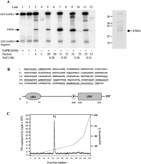
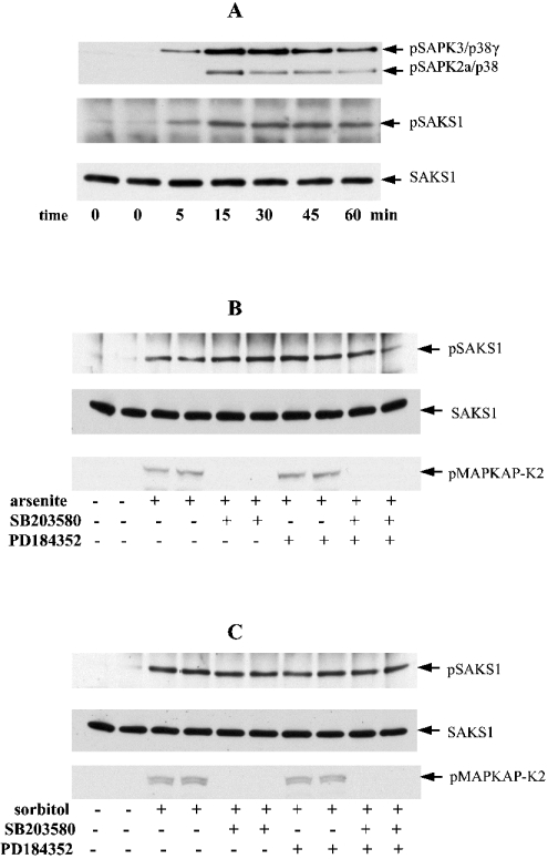
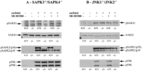
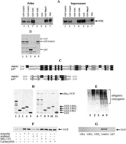
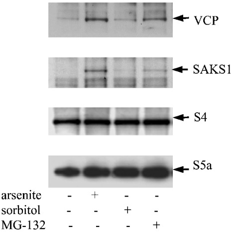
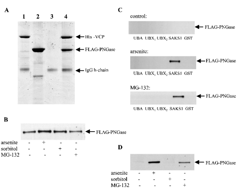
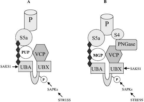
A widely expressed protein containing UBA (ubiquitin-associated) and UBX (ubiquitin-like) domains was identified as a substrate of SAPKs (stress-activated protein kinases). Termed SAKS1 (SAPK substrate-1), it was phosphorylated efficiently at Ser200 in vitro by SAPK3/p38gamma, SAPK4/p38delta and JNK (c-Jun N-terminal kinase), but weakly by SAPK2a/p38alpha, SAPK2b/p38beta2 or ERK (extracellular-signal-regulated kinase) 2. Ser200, situated immediately N-terminal to the UBX domain, became phosphorylated in HEK-293 (human embryonic kidney) cells in response to stressors. Phosphorylation was not prevented by SB 203580 (an inhibitor of SAPK2a/p38alpha and SAPK2b/p38beta2) and/or PD 184352 (which inhibits the activation of ERK1 and ERK2), and was similar in fibroblasts lacking both SAPK3/p38gamma and SAPK4/p38delta or JNK1 and JNK2. SAKS1 bound ubiquitin tetramers and VCP (valosin-containing protein) in vitro via the UBA and UBX domains respectively. The amount of VCP in cell extracts that bound to immobilized GST (glutathione S-transferase)-SAKS1 was enhanced by elevating the level of polyubiquitinated proteins, while SAKS1 and VCP in extracts were coimmunoprecipitated with an antibody raised against S5a, a component of the 19 S proteasomal subunit that binds polyubiquitinated proteins. PNGase (peptide N-glycanase) formed a 1:1 complex with VCP and, for this reason, also bound to immobilized GST-SAKS1. We suggest that SAKS1 may be an adaptor that directs VCP to polyubiquitinated proteins, and PNGase to misfolded glycoproteins, facilitating their destruction by the proteasome.


Figures

Figure 1
Figure 1. Characterization of the 43 kDa substrate of SAPK3/p38γ as SAKS1
(A) Skeletal muscle extracts were chromatographed on SP-Sepharose, fractionated from 18 to 24% PEG and chromatographed on Source 15 S. The fractions containing the 43 kDa substrate of SAPK3/p38γ were pooled and applied to heparin–Sepharose (see the Materials and methods section). Aliquots of each fraction (1 μl) were phosphorylated for 5 min at 30 °C in a 0.03 ml incubation with 2 mM MnCl2 and 20 nM [γ-32P]ATP (4×10⁶ c.p.m./nmol) in the presence or absence of SAPK3/p38γ. Reactions were terminated in SDS, subjected to SDS/PAGE, transferred on to PVDF membranes and autoradiographed. Lanes 1 and 2 show autophosphorylation of GST–SAPK3/p38γ, and lanes 3 and 4 show the pooled fractions from Source 15 S that were applied to heparin–Sepharose (L, load). Lanes 5–12 are fractions from the heparin–Sepharose column containing the 43 kDa substrate, which were incubated with MnATP in the presence (+) or absence (−) of SAPK3/p38γ. The positions of autophosphorylated GST–SAPK3/p38γ, a small proteolytic fragment of GST–SAPK3/p38γ, and the 43 kDa substrate are marked. An aliquot of fraction 21 was also subjected to SDS/PAGE and stained with SYPRO orange (right-hand panel). (B) Amino acid sequence and domain structure of the 43 kDa protein from (A), termed SAKS1. Peptides identified by MS (broken lines) or Edman sequencing (solid line) and the phosphorylation site Ser200 (*) are indicated. (C) Identification of the phosphorylation site on SAKS1. SAKS1 from heparin–Sepharose was phosphorylated for 60 min at 30 °C with SAPK3/p38γ and 0.1 mM [γ-32P]ATP (106 c.p.m./nmol) and subjected to SDS/PAGE as in (A). The phosphorylated band was excised, digested with trypsin, and the peptides were separated by chromatography on a Vydac C18 column equilibrated in 0.1% (v/v) TFA (trifluoroacetic acid). The column was developed with an acetonitrile gradient in 0.1% TFA (broken line), and fractions of 0.4 ml were collected. The site of phosphorylation in the single 32P-labelled peptide T1 (solid line) was identified as described in the text.
Figure 2
Figure 2. Activation of SAPK3/p38γ and SAPK2a/p38, and phosphorylation of SAKS1 at Ser200, induced by stress in HEK-293 cells
(A) Cells were exposed to an osmotic shock (0.5 M sorbitol) for the times indicated. SAKS1 and SAPK3/p38γ were immunoprecipitated from the cell extracts (see the Materials and methods section) with antibodies coupled to protein-G–Sepharose. After washing the beads, bound proteins were denatured in SDS and subjected to SDS/PAGE on 10% gels. After transfer on to nitrocellulose, the membranes were immunoblotted with an antibody that recognizes the phosphorylated forms of both SAPK3/p38γ (pSAPK3/p38γ) and SAPK2/p38 (pSAPK2a/p38), an antibody that recognizes the phosphorylated form of SAKS1 (pSAKS1) and an antibody that recognizes phosphorylated and unphosphorylated SAKS1 equally well (SAKS1). pSAPK2/p38 is present because the anti-SAPK3/p38γ antibody immunoprecipitates some of this enzyme. (B, C) HEK-293 cells were incubated for 1 h without (−) or with (+) 10 μM SB 203580 and/or 2 μM PD 184352, then exposed for 60 min to a chemical stress, 0.5 mM sodium arsenite (B), or for 15 min to 0.5 M sorbitol (C). The cell lysates were then immunoblotted as in (A). A further aliquot of cell lysate (50 μg of protein) was immunoblotted (without prior immunoprecipitation) using an antibody that recognizes MAPKAP-K2 (MAP-kinase-activated protein kinase 2) phosphorylated at Thr334 (pMAPKAP-K2).
Figure 3
Figure 3. Osmotic-shock-induced phosphorylation of SAKS1 is not attenuated in fibroblasts that do not express either SAPK3/p38γ and SAPK4/p38δ or JNK1 and JNK2
(A) Primary mouse fibroblasts were isolated from day-13.5 embryos of wild-type (+/+) mice and mice that do not express SAPK3/p38γ and SAPK4/p38δ (−/−). The fibroblasts were cultured [14] and incubated for 1 h with or without 10 μM SB 203580, then exposed for 30 min to 0.5 M sorbitol. Following cell lysis, SAKS1 was immunoprecipitated from 2 mg of cell-lysate protein, subjected to SDS/PAGE and immunoblotted for the phosphorylation of SAKS1 at Ser200 and with antibodies that recognize the phosphorylated forms of SAPK3/p38γ, SAPK2/p38 and JNK. Antibodies used were against the phosphorylated form of SAKS1 (pSAKS1), both the phosphorylated and unphosphorylated SAKS1 equally (SAKS1), the phosphorylated forms of both SAPK3/p38γ (pSAPK3/p38γ) and SAPK2/p38 (pSAPK2/p38), and the phosphorylated JNK. (B) As in (A), except that immortalized fibroblasts from wild-type mice (+/+) and mice that do not express JNK1 and JNK2 (−/−) were used.
Figure 4
Figure 4. SAKS1 binds ubiquitin tetramers and VCP
(A) Full-length GST–SAKS1 (lanes 1 and 2), a fragment containing the UBA domain, GST–UBA (3–41) (lane 3), a fragment containing the UBX domain, GST–UBX1 (206–293) (lane 4), GST (lane 5) or GST–Rhp23 (lane 6) each at 0.2 μM were coupled individually to 20 μl of glutathione–Sepharose beads, then incubated with 0.05 μg of tetra-ubiquitin (Ub4) in 0.05 ml 50 mM Tris/HCl, pH 7.5, 50 mM NaCl, 1% (w/v) Triton X-100, 10% (v/v) glycerol and 0.1 mg/ml BSA. After mixing for 30 min at 4 °C and centrifugation, the supernatant was retained and the beads washed three times. The supernatant and bound material were denatured in SDS, electrophoresed on a 4–12% polyacrylamide gradient gel, transferred on to nitrocellulose membranes and immunoblotted for the presence of ubiquitin. Lane 7, 0.05 μg of Ub4 control. The GST–Rhp23 was included as a protein known to bind polyubiquitin [23]. (B) GST–SAKS1 (lanes 1 and 2), GST (lane 3), each at 10 μg, or buffer (lane 4) were coupled to glutathione–Sepharose beads and incubated for 1 h at 4 °C with 4 mg of HEK-293 cell-lysate protein. The suspensions were centrifuged, the supernatants were discarded, and the beads were washed twice in lysis buffer containing 0.25 M NaCl and twice in lysis buffer alone. Samples were denatured in SDS, electrophoresed on a 4–12% polyacrylamide gel and stained with colloidal Coomassie Blue (Invitrogen). Lane 5 is purified GST–SAKS1 and the 97 kDa protein (VCP) binding specifically to GST–SAKS1 is indicated. (C) The UBX domains of SAKS1 and p47 are compared with identities highlighted in black and conservative replacements in grey. (D) A 10 μg volume of bacterially expressed full-length GST–SAKS1 (lane 1), GST–UBA (3–41) (lane 2), GST–UBX1 (206–293) (lane 3), GST–UBX2 (180–297) (lane 4) or GST (lane 6) was denatured in SDS, electrophoresed on a 4–12% polyacrylamide gradient gel and stained with colloidal Coomassie Blue. A volume of 10 μg of bacterially expressed full-length GST–SAKS1 (lanes 6 and 7), GST–UBA (3–41) (lane 8) GST–UBX1 (206–293) (lane 9), GST–UBX2 (180–297) (lane 10) or GST (lane 11) were coupled to 20 μl of glutathione–Sepharose in 0.5 ml of 50 mM Tris/HCl, pH 7.5, 0.15 M KCl, 1 mM DTT, 1 mM MgCl2, 1 mM ATP and 0.03% (w/v) Brij 35. After washing the beads, 10 μg of His6–VCP in 0.5 ml of buffer was added and the suspension was mixed end-over-end for 30 min at 21 °C. After centrifugation for 1 min at 13000 g, the supernatant was removed, the beads were washed, and attached proteins were subjected to SDS/PAGE as in (B). Protein staining bands migrating more rapidly than GST–SAKS1 (lanes 1 and 2) are proteolytic fragments truncated at the C-terminus. (E) HEK-293 cells were left unstimulated (lane 1), exposed for 1 h to 0.5 mM sodium arsenite (lane 2), incubated for 30 min with 0.5 M sorbitol (lane 3) or incubated for 3 h with 20 μM MG-132 (lane 4) or 20 μM lactacystin (lane 5). Following cell lysis, 15 μg of lysate protein was subjected to SDS/PAGE and immunoblotted with an anti-ubiquitin antibody. (F) Cells were left unstimulated or exposed to sodium arsenite, sorbitol, MG-132 or lactacystin as in (E). Following cell lysis, 3 mg of lysate protein was incubated with 2 μg of GST–SAKS1 bound to glutathione–Sepharose, and the VCP bound to glutathione–Sepharose was analysed by immunoblotting. (G) Cells were incubated with sodium arsenite as in (B) and the VCP associated with full-length GST–SAKS1, GST–UBA, GST–UBX1 (206–293), GST–UBX2 (180–297) and GST were immunoblotted as in (F).
Figure 5
Figure 5. SAKS1, VCP and S4 are present in anti-S5a immunoprecipitates
HEK-293 cells were incubated without (−) or with (+) sodium arsenite, sorbitol or MG-132, as in Figure 4(E), and lysed. The S5a protein was immunoprecipitated from 3 mg of cell-lysate protein, and the immunoprecipitates were analysed for the presence of VCP, SAKS1, S4 and S5a by immunoblotting.
Figure 6
Figure 6. PGNase forms a complex with VCP and therefore interacts indirectly with SAKS1
(A) Bacterially expressed His6–VCP (2.5 μg) was immunoprecipitated in 0.5 ml of the buffer used in Figure 4(D) using an anti-His6 antibody coupled to protein-G–Sepharose. The supernatant was discarded, the beads were washed once with 1 ml of buffer, and incubated without (lane 1) or with (lane 4) a several-fold molar excess of FLAG–PNGase in 0.5 ml of buffer. The suspensions were mixed end-over-end for 30 min at 21 °C, and the beads were collected and washed as before. After denaturation in SDS, the solubilized proteins were electrophoresed on 4–12% polyacrylamide gradient gels, stained with colloidal Coomassie Blue and destained. Lane 2, same as lane 1 except that His6–VCP was omitted; lane 3, same as lane 1 except that His6–VCP and anti-His6 antibody were both omitted. The position of the IgG heavy (h)-chain is indicated. (B) Vectors expressing FLAG–PNGase and His6–VCP were transfected into HEK-293 cells. The cells were then either left untreated (control) or incubated with sodium arsenite, or MG-132 as in Figures 4(E) and 4(F). The cells were lysed and 1 mg of lysate protein was incubated for 1 h with 0.5 μg of anti-His6 antibody bound to protein-G–Sepharose. The Sepharose beads were washed and immunoblotted with an anti-FLAG antibody as in (B). (C) Vectors expressing FLAG–PNGase were transfected into HEK-293 cells. The cells were then incubated with sodium arsenite, sorbitol or MG-132 as in Figures 4(E) and 4(F). The cells were lysed and 3 mg of lysate protein was incubated with 2 μg of GST, GST–SAKS1 or the GST–SAKS1 fragments used in Figure 4(G), each bound to glutathione–Sepharose. The PNGase bound to glutathione–Sepharose was then analysed by immunoblotting with an anti-FLAG antibody. (D) The endogenous SAKS1 in cell extracts was immunoprecipitated from 2 mg of cell-lysate protein and the amount of FLAG–PNGase present in the immunoprecipitates was analysed by immunoblotting.
Figure 7
Figure 7. Schematic representation of the proposed role of SAKS1 in localizing VCP to ubiquitinated proteins and PNGase to MGPs
The four black diamonds denote a polyubiquitin chain, which binds to the UBA domain of SAKS1. (A) VCP is directed to polyubiquitinated proteins (PUPs) via the adaptor protein SAKS1, allowing VCP to unfold PUPs in an ATP-requiring process. PUPs can then be degraded by the proteasome with which they interact via the S5a subunit of the 19 S proteasomal subunit (P). (B) PNGase is directed to polyubiquitinated MGPs via VCP and the adaptor protein SAKS1, allowing PNGase to deglycosylate MGPs, which can then be degraded by the proteasome. PNGase itself is reported to bind to the S4 component of the 19 S proteasome.

References

    1. Fukunaga R., Hunter T. MNK1, a new MAP kinase-activated protein kinase, isolated by a novel expression screening method for identifying protein kinase substrates. EMBO J. 1997;16:1921–1933. - PMC - PubMed
    1. McLaughlin M. M., Kumar S., McDonnell P. C., Van Horn S., Lee J. C., Livi G. P., Young P. R. Identification of mitogen-activated protein (MAP) kinase-activated protein kinase-3, a novel substrate of CSBP p38 MAP kinase. J. Biol. Chem. 1996;271:8488–8492. - PubMed
    1. Balendran A., Casamayor A., Deak M., Paterson A., Gaffney P., Currie R., Downes C. P., Alessi D. R. PDK1 acquires PDK2 activity in the presence of a synthetic peptide derived from the carboxyl terminus of PRK2. Curr. Biol. 1999;22:393–404. - PubMed
    1. New L., Jiang Y., Zhao M., Liu K., Zhu W., Flood L. J., Kato Y., Parry G. C., Han J. PRAK, a novel protein kinase regulated by the p38 MAP kinase. EMBO J. 1998;17:3372–3384. - PMC - PubMed
    1. Yaffe M. B., Lebarc G. G., Lai J., Obata T., Volinia S., Cantley L. C. A motif-based profile scanning approach for genome-wide prediction of signaling pathways. Nat. Biotechnol. 2001;19:348–353. - PubMed

Publication types

MeSH terms