. 2005 Jan 15;385(Pt 2):427-32.
doi: 10.1042/BJ20041218.

The high concentration of Arg213-->Gly extracellular superoxide dismutase (EC-SOD) in plasma is caused by a reduction of both heparin and collagen affinities

Affiliations

The high concentration of Arg213-->Gly extracellular superoxide dismutase (EC-SOD) in plasma is caused by a reduction of both heparin and collagen affinities

Steen V Petersen et al. Biochem J. .

Abstract

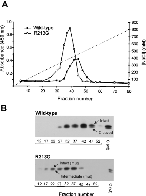
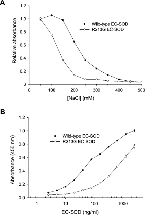
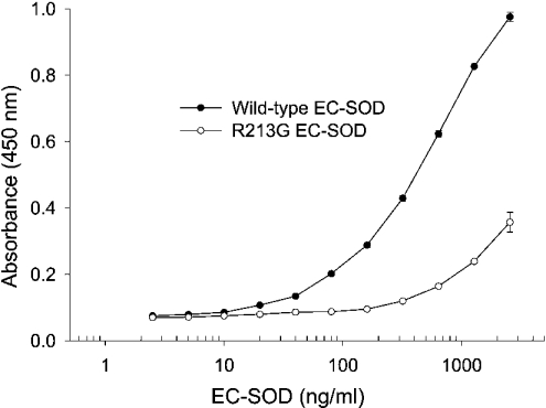
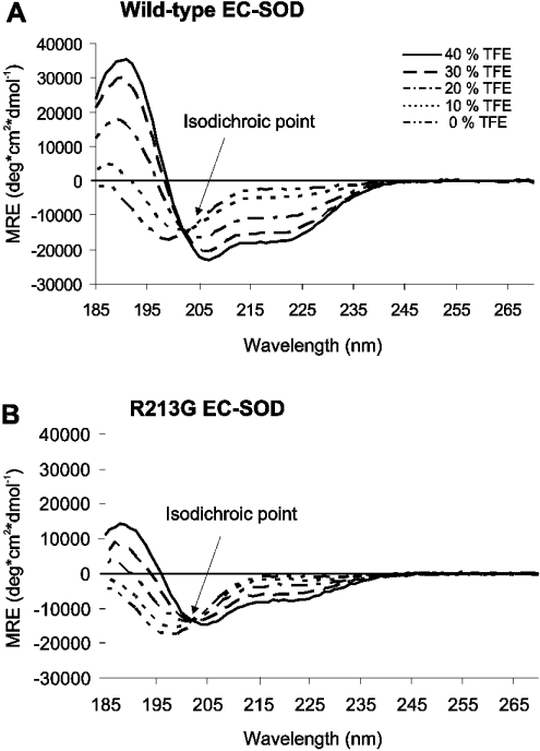
The C-terminal region of EC-SOD (extracellular superoxide dismutase) mediates the binding to both heparin/heparan sulphate and type I collagen. A mutation (Arg213-->Gly; R213G) within this extracellular matrix-binding region has recently been implicated in the development of heart disease. This relatively common mutation affects the heparin affinity, and the concentration of EC-SOD in the plasma of R213G homozygous individuals is increased 10- to 30-fold. In the present study we confirm, using R213G EC-SOD purified from a homozygous individual, that the heparin affinity is reduced. Significantly, the collagen affinity of the R213G EC-SOD variant was similarly affected and both the heparin and collagen affinities were reduced by 12-fold. Structural analysis of synthetic extracellular matrix-binding regions suggests that the mutation alters the secondary structure. We conclude that the increased concentration of EC-SOD in the plasma of R213G carriers is caused by a reduction in both heparin and collagen affinities.


Figures

Figure 1
Figure 1. Affinity purification of EC-SOD
(A) Plasma from a wild-type individual (25 ml) and a homozygous R213G individual (5 ml) was applied to a heparin–Sepharose column and bound proteins were subsequently eluted with NaCl (indicated by broken line). Collected fractions were analysed for the presence of EC-SOD by using a heparin-based ELISA assay. The amount of EC-SOD is given as the absolute absorbance. (B) Collected fractions were analysed by SDS/PAGE using 600 and 40 μl of fractions representing wild-type and R213G plasma respectively. EC-SOD was subsequently detected by using Western blotting. The intact, intermediate and cleaved forms of EC-SOD are indicated. C, wild-type EC-SOD purified from human aorta used as control.
Figure 2
Figure 2. Heparin-binding analysis of EC-SOD
(A) Approximately 100 ng/ml of wild-type and R213G EC-SOD were allowed to bind a heparin–BSA-coated surface in the presence of increasing concentrations of NaCl. For comparison, the binding of EC-SOD is depicted as the absorbance relative to the binding in the presence of 50 mM NaCl. (B) Wild-type and R213G EC-SOD were diluted to the indicated concentrations in a buffer containing 135 mM NaCl. The qualitative amount of bound material is given as absorbance at 450 nm. The experiment was repeated three times producing similar results. The data points represent the mean for doublet estimations and error bars indicate S.D.
Figure 3
Figure 3. Collagen-binding capacity of native EC-SOD
Microtitre wells were coated with type I collagen and subsequently incubated with wild-type and R213G EC-SOD at the concentrations indicated. The R213G variant was purified from a homozygous individual. The amount of bound material is given as absorbance at 450 nm. The experiment was repeated three times producing similar results. The data points represent the mean for doublet estimations and error bars indicate S.D.
Figure 4
Figure 4. Structural analysis of homodimeric peptides
Far-UV CD spectra of (A) wild-type and (B) R213G homodimeric peptides were recorded at 20 °C. Peptides were analysed at 0.2 mg/ml in 3 mM NaPO4 (pH 7.4) in the presence of increasing amounts of TFE, as indicated. Local peaks at 190, 208 and 222 nm indicate the presence of α-helical structures. The isodichroic point at 203 nm indicates a two-state process.

References

    1. Fattman C. L., Schaefer L. M., Oury T. D. Extracellular superoxide dismutase in biology and medicine. Free Radical Biol. Med. 2003;35:236–256. - PubMed
    1. Marklund S. L. Human copper-containing superoxide dismutase of high molecular weight. Proc. Natl. Acad. Sci. U.S.A. 1982;79:7634–7638. - PMC - PubMed
    1. Oury T. D., Crapo J. D., Valnickova Z., Enghild J. J. Human extracellular superoxide dismutase is a tetramer composed of two disulphide-linked dimers: a simplified, high-yield purification of extracellular superoxide dismutase. Biochem. J. 1996;317:51–57. - PMC - PubMed
    1. Petersen S. V., Oury T. D., Valnickova Z., Thogersen I. B., Hojrup P., Crapo J. D., Enghild J. J. The dual nature of human extracellular superoxide dismutase: one sequence and two structures. Proc. Natl. Acad. Sci. U.S.A. 2003;100:13875–13880. - PMC - PubMed
    1. Karlsson K., Lindahl U., Marklund S. L. Binding of human extracellular superoxide dismutase C to sulphated glycosaminoglycans. Biochem. J. 1988;256:29–33. - PMC - PubMed

Publication types

MeSH terms