. 2004 Sep 13;166(6):801-13.
doi: 10.1083/jcb.200405128.

Functional interaction between BLM helicase and 53BP1 in a Chk1-mediated pathway during S-phase arrest

Affiliations

Functional interaction between BLM helicase and 53BP1 in a Chk1-mediated pathway during S-phase arrest

Sagar Sengupta et al. J Cell Biol. .

Abstract

Bloom's syndrome is a rare autosomal recessive genetic disorder characterized by chromosomal aberrations, genetic instability, and cancer predisposition, all of which may be the result of abnormal signal transduction during DNA damage recognition. Here, we show that BLM is an intermediate responder to stalled DNA replication forks. BLM colocalized and physically interacted with the DNA damage response proteins 53BP1 and H2AX. Although BLM facilitated physical interaction between p53 and 53BP1, 53BP1 was required for efficient accumulation of both BLM and p53 at the sites of stalled replication. The accumulation of BLM/53BP1 foci and the physical interaction between them was independent of gamma-H2AX. The active Chk1 kinase was essential for both the accurate focal colocalization of 53BP1 with BLM and the consequent stabilization of BLM. Once the ATR/Chk1- and 53BP1-mediated signal from replicational stress is received, BLM functions in multiple downstream repair processes, thereby fulfilling its role as a caretaker tumor suppressor.


Figures
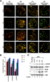
Figure 1.
Figure 1.
γ-H2AX, 53BP1, BLM, and p53 colocalize and accumulate with similar kinetics. (A) Time course of the accumulation of BrdU, γ-H2AX, 53BP1, BLM, and p53 foci after HU treatment. NHFs were contact inhibited, split at low dilution, and treated with HU for the indicated time intervals. BrdU staining was done on washed cells and incubated with the nucleotide analogue for 10 min. Immunofluorescence was performed with antibodies against BrdU/γ-H2AX (a); 53BP1 (monoclonal)/γ-H2AX (b); 53BP1 (monoclonal)/BLM (ab476) (c); and 53BP1 (monoclonal)/p53 (CM1) (d). Bars, 5 μm. (B) Quantitation of A. The graphs represent mean ± SD. (C) The accumulation of proteins after HU treatment. Western blots were performed with anti-γ-H2AX (a), anti-53BP1 (polyclonal; b), anti-BLM (C-18) (c), anti-p53 (DO-1) (d), and anti-TBP antibodies (e).
Figure 2.
Figure 2.
53BP1 interacts with p53 via BLM. (A and C) The immunoprecipitation of BLM revealed 53BP1 in NHFs and NHF E6. NHFs (A) and NHF E6 (C) were either left asynchronous (lane 1) or contact inhibited, split at low dilution, and treated with HU (lane 2) for 6 h. 400 μg of lysates were immunoprecipitated with antibodies against BLM (C-18) in the absence (lanes 3 and 4) or presence (lane 5) of the blocking peptide. Input (lanes 1 and 2) indicates 10% of the lysate was used for immunoprecipitation. The efficiency of immunoprecipitation was verified with self-antibody (a). The BLM immunoprecipitates were probed with anti-53BP1 (polyclonal; for both A and C, b) and anti-p53 (DO-1) (for A only, c) antibodies. Lanes 1 and 3, minus HU; lanes 2, 4, and 5, plus HU. (B and D) Immunoprecipitation of p53 revealed 53BP1 in NHFs, but not in BS. Same as in A and C, except NHFs were used in B and BS in D. Immunoprecipitation was done with antibodies against p53 (DO-1) (lanes 3 and 4) or against the corresponding IgG (lane 5). The p53 immunoprecipitates were probed with polyclonal anti-53BP1 antibody (b). (E) γ-H2AX, 53BP1, and BLM colocalize in the absence of p53. NHFs or NHF E6 were prepared as in A and treated with HU for 6 h. Immunofluorescence was performed with antibodies against γ-H2AX/53BP1 (monoclonal; a) and BLM (ab476)/53BP1 (monoclonal; b). Bars, 5 μm. (F) 53BP1 accumulates at the sites of stalled replication forks even in the absence of BLM. Same as in E except, BS and BS A-15 cells were used. Bars, 5 μm.
Figure 3.
Figure 3.
53BP1 regulates the accumulation of BLM. (A) Absence of 53BP1 leads to nucleolar accumulation of BLM in KO MEFs. WT or 53BP1 KO MEFs were treated with HU for 6 h. Immunofluorescence was performed with antibodies against BLM (ab476)/53BP1 (monoclonal; a); p53 (CM1)/53BP1 (monoclonal; b); and BLM (ab476)/nucleolin (c). (B) The interaction of BLM with 53BP1 and p53 is decreased in 53BP1siRNA-treated cells. Control (lanes 1 and 2) or 53BP1 siRNA-transfected (lanes 3 and 4) NHFs were contact inhibited, split at low dilution, and grown either in the absence (lanes 1 and 3) or presence (lanes 2 and 4) of HU for 6 h. 400 μg of lysates were immunoprecipitated with antibodies against BLM (C-18) (lanes 5–9) in the absence (lanes 5–8) or presence (lane 9) of the blocking peptide. Input (lanes 1–4) indicates 10% of the lysate was used for immunoprecipitation. The efficiency of immunoprecipitation was verified with BLM (ab476) antibody (a). Antibodies used for the detection of other proteins in BLM immunoprecipitation were anti-53BP1 (polyclonal; b and d) and anti-p53 (DO-1) (c and e). Lanes 1, 3, 5, and 7, minus HU; lanes 2, 4, 6, 8, and 9, plus HU. Panels d and e are longer exposures of the same blots in b and c. (C) The lack of 53BP1 prevents the accumulation of BLM and p53. Transfection and treatment were done as in B. Immunofluorescence was performed with antibodies against BLM (ab476)/53BP1 (monoclonal)/DNA (DAPI) (a) and p53 (CM1)/53BP1 (monoclonal)/DNA (DAPI) (b). Fields were chosen that contained a cell in which 53BP1 expression was not suppressed as an internal control for cells lacking expression of 53BP1. Arrows indicate the position of cells lacking 53BP1 expression. (D) Quantitation of C. The histogram represents mean ± SD.
Figure 4.
Figure 4.
53BP1 and BLM can accumulate and physically interact independently of γ-H2AX. (A) The accumulation of proteins in WT and H2AX KO MEFs after HU. H2AX WT (lanes 1 and 2) and KO (lanes 3 and 4) cells were contact inhibited, split at low dilution, and grown either in the absence (lanes 1 and 3) or presence (lanes 2 and 4) of HU for 6 h. Western blots were performed with anti-53BP1 (polyclonal; a), anti-BLM (ab476) (b), anti-γ-H2AX (c), and anti-TBP antibodies (d). (B) 53BP1 and BLM accumulate at the sites of stalled replication forks even in the absence of γ-H2AX. MEFs were treated as in A. Immunofluorescence was performed with antibodies against γ-H2AX /53BP1 (monoclonal; a) and BLM (ab476)/53BP1 (monoclonal; b). Bars, 5 μm. (C) Quantitation of B. The histogram represent mean ± SD. (D) BLM interacts with 53BP1 and BLM even in the absence of H2AX. WT (lanes 1 and 2) or H2AX KO (lanes 3 and 4) MEFs were prepared as in A and were either left untreated (lanes 1 and 3) or treated with HU (lane 2 and 4) for 6 h. 400 μg of lysates were immunoprecipitated with antibodies against BLM (ab476) (lanes 5–8) or corresponding IgG (lane 9). Input (lanes 1–4) indicates 10% of the lysate used for immunoprecipitation. The efficiency of immunoprecipitation was verified with BLM (ab476) antibody (a). Antibodies used for the detection of other proteins in BLM immunoprecipitation were anti-53BP1 (polyclonal) (b) and γ-H2AX (c). Lanes 1, 3, 5, 7, minus HU; lanes 2, 4, 6, 8, and 9, plus HU.
Figure 5.
Figure 5.
Accumulation of 53BP1 and BLM depends on the presence of Chk1 and ATR. (A) The accumulation of proteins in control and Chk1 siRNA-treated NHFs. Control (lanes 1 and 2) or Chk1 siRNA-transfected (lanes 3 and 4) NHFs were contact inhibited, split at low dilution, and grown either in the absence (lanes 1 and 3) or presence (lanes 2 and 4) of HU for 6 h. Western blots were performed with anti-Chk1 (a), anti-pChk1 (b), anti-53BP1 (polyclonal; c), anti-BLM (C-18) (d), and anti-TBP antibodies (e). (B) The lack of Chk1 prevents the accumulation of pChk1, BLM, and p53. NHFs transfection and treatment was done as in A. Immunofluorescence was performed with antibodies against BLM (C-18)/pChk1/DNA (DAPI) (a) and 53BP1 (monoclonal)/pChk1/DNA (DAPI) (b). Fields were chosen that contained a cell in which Chk1 expression was not suppressed as an internal control for the cells lacking expression of Chk1. Arrows indicate position of cells that lack the expression of Chk1. Bars, 5 μm. (C) Quantitations of Fig. 5 (B, E, and G). The histograms represent mean ± SD. (D) The accumulation of proteins in NHFs due to treatment with UCN-01 and the transfection of Chk1 wild-type (WT) or antisense (AS). NHFs, transfected with Chk1 WT (lane 3) or AS (lane 4) were contact inhibited, split at low dilution, and either left untreated (lane 1) or treated with HU (lanes 2–4). Untransfected HU-treated cells were coincubated with UCN-01 (lane 5). Western blots were performed with anti-Chk1 (a), anti-pChk1 (b), anti-BLM (C-18) (c), anti-53BP1 (polyclonal; d), and anti-TBP antibodies (e). (E) UCN-01 inhibits focal colocalization of BLM/53BP1 with pChk1. NHFs were prepared as in D and treated with HU (a) or HU and UCN-01 (b) for 6 h. Immunofluorescence was performed with antibodies against pChk1/BLM (C-18) or 53BP1 (monoclonal)/BLM (ab476). (F) The accumulation of proteins in NHFs after treatment with caffeine and LY294002. NHFs were contact inhibited, split at low dilution, and left either untreated (lane 1) or treated (lanes 2–4) with HU. Coincubation was performed with either caffeine (lane 3) or LY294002 (lane 4). Western blots were performed with anti-pChk1 (a), anti-53BP1 (polyclonal; b), anti-BLM (C-18) (c), and anti-TBP (d) antibodies. Bars, 5 μm. (G) Caffeine and LY294002 inhibit focal colocalization of 53BP1 and BLM. NHFs were prepared as in F and were treated with HU (a); HU and caffeine (b); and HU and LY294002 (c) for 6 h. Immunofluorescence was performed with antibodies against pChk1/BLM (C-18) or 53BP1 (monoclonal)/BLM (ab476). Bars, 5 μm.
Figure 6.
Figure 6.
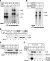
Chk1-mediated phosphorylation of BLM. (A) Chk1 phosphorylates p53 and BLM. Recombinant Chk1 (WT or KD) was incubated with full-length p53 or BLM protein in the presence of γ-32P ATP. The proteins were resolved by SDS-PAGE and detected by autoradiography. (B) Chk1 phosphorylates BLM T99A/T122A mutant. Phosphorylation reactions were performed as in A except immunoprecipitated WT BLM or T99A/T122A mutant was used. (C) Chk1 and BLM interact in vivo. NHFs were contact inhibited, split at low dilution, and treated with HU for 6 h. Lysates (2 mg) were immunoprecipitated with antibodies against BLM (ab476) (lanes 3 and 4), Chk1 (lanes 6 and 7), or the corresponding goat IgGs (lane 5, goat; lane 8, rabbit). Input (lanes 1 and 2) indicates 5% of the lysate was used for immunoprecipitation. The blots were probed with BLM (ab476) (a) and Chk1 (b) antibodies. Lanes 1, 3, 6, minus HU; lanes 2, 4, 5, 7, and 8, plus HU. (D) Inhibition of Chk1-mediated phosphorylation destabilizes BLM. NHFs were either left untreated (lane 1) or treated with HU (lane 2), LLnL (lane 3), HU+UCN-01 (lane 4), and HU+UCN-01+LLnL (lane 5). Western blots were performed with anti-BLM (C-18) (a) and anti-TBP (b) antibodies. (E) Chk1-mediated phosphorylation of BLM on serine residues in vivo. NHFs were treated as in D (for a and b) or grown in the presence of phosphate-free DME containing 2 mCi of 32P-labeled inorganic phosphate per ml (for c). Lysates (2 mg) were immunoprecipitated with antibodies against BLM (ab476) (lanes 1–5) or the corresponding IgG (lane 6). The efficiency of immunoprecipitation was verified with (a) BLM (ab476) antibody. Phosphorylation on BLM was detected with phosphoserine antibody (b) or by autoradiography (c).

References

    1. Abraham, R.T. 2001. Cell cycle checkpoint signaling through the ATM and ATR kinases. Genes Dev. 15:2177–2196. - PubMed
    1. Anderson, L., C. Henderson, and Y. Adachi. 2001. Phosphorylation and rapid relocalization of 53BP1 to nuclear foci upon DNA damage. Mol. Cell. Biol. 21:1719–1729. - PMC - PubMed
    1. Bartek, J., and J. Lukas. 2003. Chk1 and Chk2 kinases in checkpoint control and cancer. Cancer Cell. 3:421–429. - PubMed
    1. Bassing, C.H., K.F. Chua, J. Sekiguchi, H. Suh, S.R. Whitlow, J.C. Fleming, B.C. Monroe, D.N. Ciccone, C. Yan, K. Vlasakova, et al. 2002. Increased ionizing radiation sensitivity and genomic instability in the absence of histone H2AX. Proc. Natl. Acad. Sci. USA. 99:8173–8178. - PMC - PubMed
    1. Beamish, H., P. Kedar, H. Kaneko, P. Chen, T. Fukao, C. Peng, S. Beresten, N. Gueven, D. Purdie, S. Lees-Miller, et al. 2002. Functional link between BLM defective in Bloom's syndrome and the ataxia-telangiectasia-mutated protein, ATM. J. Biol. Chem. 277:30515–30523. - PubMed

MeSH terms