Comparative Study
. 2004 Sep 13;166(6):865-76.
doi: 10.1083/jcb.200403004.

Rb is required for progression through myogenic differentiation but not maintenance of terminal differentiation

Affiliations
Comparative Study

Rb is required for progression through myogenic differentiation but not maintenance of terminal differentiation

Michael S Huh et al. J Cell Biol. .

Abstract

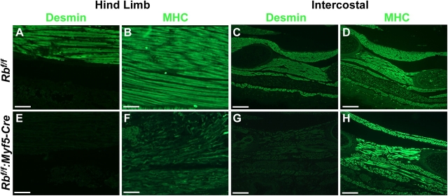
To investigate the requirement for pRb in myogenic differentiation, a floxed Rb allele was deleted either in proliferating myoblasts or after differentiation. Myf5-Cre mice, lacking pRb in myoblasts, died immediately at birth and exhibited high numbers of apoptotic nuclei and an almost complete absence of myofibers. In contrast, MCK-Cre mice, lacking pRb in differentiated fibers, were viable and exhibited a normal muscle phenotype and ability to regenerate. Induction of differentiation of Rb-deficient primary myoblasts resulted in high rates of apoptosis and a total inability to form multinucleated myotubes. Upon induction of differentiation, Rb-deficient myoblasts up-regulated myogenin, an immediate early marker of differentiation, but failed to down-regulate Pax7 and exhibited growth in low serum conditions. Primary myoblasts in which Rb was deleted after expression of differentiated MCK-Cre formed normal multinucleated myotubes that did not enter S-phase in response to serum stimulation. Therefore, Rb plays a crucial role in the switch from proliferation to differentiation rather than maintenance of the terminally differentiated state.


Figures

Figure 1.
Figure 1.
Severely impaired myogenesis in P0 Rb f/f :Myf5-Cre mice. Hematoxylin and eosin staining of paraffin-embedded longitudinal sections through the hind limb of control (A and C) and Rb f/f :Myf5-Cre (B and D) mice. Hematoxylin and eosin staining of cross sections through diaphragm of control (E) and Rb f/f :Myf5-Cre (F) mice. Bars: (A and B) 400 μm; (C–F) 50 μm.
Figure 2.
Figure 2.
Abnormal skeletal muscle differentiation in P0 Rb f/f :Myf5-Cre mice. Immunofluorescent staining of longitudinal sections through the hind limb of control (A and B) and Rb f/f :Myf5-Cre (E and F) mice with antibodies to desmin and myosin heavy chain (MHC). Immunofluorescent staining of intercostal muscles of control (C and D) and Rb f/f :Myf5-Cre (G and H) mice with antibodies to desmin and MHC. Bars: (A, B, E, and F) 50 μm; (C, D, G, and H) 100 μm.
Figure 3.
Figure 3.
Rb f/f :MCK-Cre mice have normal skeletal muscle. Hematoxylin and eosin staining of frozen cross sections of control muscles (A–C) and Rb f/f :MCK-Cre muscles (D–F). TA, tibialis anterior; EDL, extensor digitorum longus. Phase-contrast micrograph of X-Gal–stained single muscle fibers of R26R3 control (G) and R26R3:MCK-Cre (H) mice. (I) 32P end-labeled PCR genotype analysis of the floxed Rb locus revealed 98–99% excision. 32P end-labeled PCR was performed on DNA samples extracted from a pool of 20 single fibers from three independent mice (Rb f/f :MCK-Cre A, Rb f/f :MCK-Cre B, and Rb f/f :MCK-Cre C). (J) pRb immunoblot analysis of single-fiber protein extracts reveals virtually no detectable pRb. α-Tubulin protein levels were used as loading controls. Protein was extracted from a pool of 200 single fibers from Rb f/f :MCK-Cre A and Rb f/f :MCK-Cre B mice, and from Rb f/wt A and Rb f/wt B littermate controls. Protein extracts from Ad-Lac-Z– and Ad-Cre–infected Rb f/f primary myoblasts were used as positive and negative controls, respectively. Bars, 50 μm.
Figure 4.
Figure 4.
Adenoviral delivery of Cre recombinase into Rb floxed primary myoblasts completely eliminates pRb expression. (A) 32P end-labeled PCR genotype analysis of the floxed Rb locus revealed complete excision. Primers flanking the LoxP sites in the Rb locus were used to amplify DNA samples from Ad-Lac-Z control– and Ad-Cre–infected Rb flox homozygous (f/f) primary myoblasts from three independent isolates (Rb f/f A, Rb f/f B, and Rb f/f C). (B) Immunoblot detection of pRb from control- and Ad-Cre–infected primary myoblast protein extracts indicated a complete absence of pRb. Whole cell protein extract was harvested from primary myoblasts in growth media (GM) and after 1 d in DM (D1). (C) Immunofluorescent staining of control- and Ad-Cre–infected Rb f/f primary myoblasts. Infected primary myoblasts were PFA fixed after 2 d in DM and stained with antibody to pRb.
Figure 5.
Figure 5.
pRb null primary myoblasts exhibit high rates of apoptosis in response to serum withdrawal. Apoptotic cell death was assessed by fluorescein-conjugated TUNEL analysis after 6 h in DM on control- (A–C) and Ad-Cre– infected (D–F) Rb f/f primary myoblasts. Primary myoblasts were counterstained with propidium iodide (PI).
Figure 6.
Figure 6.
pRb null primary myoblasts are incapable of forming terminally differentiated multinucleated myotubes. Double immunofluorescent staining of control- (A–C) and Ad-Cre–infected (D–F) Rb f/f primary myoblasts with antibodies to MyoD (FITC) and MHC (rhodamine). Immunofluorescent staining of control- (G–I) and Ad-Cre–infected (J–L) Rb f/f myoblasts with antibody to myogenin (rhodamine). Nuclei were counterstained with DAPI. Infected primary myoblasts were PFA fixed and stained at the indicated time points. GM, growth; DM D1, differentiation day 1; DM D5, differentiation day 5.
Figure 7.
Figure 7.
Absence of terminal differentiation in pRb null primary myoblasts correlates with an inability to down-regulate markers of proliferation and early markers of myogenic differentiation. (A) RNase protection assay for cyclinA2 and cyclinB1 mRNA in control- and Ad-Cre–infected Rb f/f primary myoblasts. (B) RNase protection assay for cdk1, cdk2, and cdk4 mRNA in control- and Ad-Cre–infected Rb f/f myoblasts. GAPDH mRNA levels were used as loading controls. Total RNA was harvested from infected myoblasts at the indicated time points (A and B). GM, growth; DM 36hr, differentiation 36 h. (A and B) White lines indicate that intervening lanes have been spliced out. (C) Immunoblot differentiation time course analysis of p107, p130, Pax7, MyoD, myogenin, and MHC in control- and Ad-Cre–infected Rb f/f myoblasts. α-Tubulin protein levels were used as loading controls. Whole cell protein extract was harvested at the indicated time points. GM, growth; D1, differentiation day 1; D3, differentiation day 3; D5, differentiation day 5. Protein extract from 293T cells was used as a nonmyogenic control.
Figure 8.
Figure 8.
Serum restimulation is unable to induce DNA synthesis in terminally differentiated Rb f/f :MCK-Cre myotubes. Double immunofluorescent staining for BrdU (FITC) and MHC (rhodamine) in Rb f/f :Ad-Lac-Z–infected (A and B), Rb f/f :Ad-Cre–infected (C and D), and Rb f/f :MCK-Cre (E and F) primary myoblasts. Primary myoblasts were cultured in DM for 4 d and were either BrdU pulsed for 1 h before fixation (DM D4) or restimulated with BrdU-supplemented growth media for 24 h before fixation (DM D4 + GM 24 h).
Figure 9.
Figure 9.
MCK-Cre transgene efficiently eliminates pRb expression in Rb f/ f:MCK-Cre differentiated primary myotubes. (A) 32P end- labeled PCR genotype analysis of the floxed Rb locus. Primers flanking the LoxP sites in the Rb locus were used to amplify DNA samples from Rb f/f :MCK-Cre primary myoblasts during growth (GM) and after 5 d in differentiation conditions (D5) in three independent isolates (Rb f/f :MCK-Cre 1–3). (B) Immunoblot detection of pRb from Rb wt/wt :MCK-Cre control and Rb f/f :MCK-Cre primary myoblast protein extracts. α-Tubulin protein levels were used as loading controls. Whole cell protein extract was harvested from primary myoblasts in growth media (GM) and after 5 d in DM (D5). (C) Immunofluorescent staining of Rb wt/wt :MCK-Cre control and Rb f/f :MCK-Cre primary myotubes. Myotubes were PFA fixed after 5 d in DM, stained with antibodies to pRb (FITC) and MHC (rhodamine), and counterstained with DAPI.
Figure 10.
Figure 10.
Late-stage removal of pRb in differentiated Rb f/ f:MCK-Cre myotubes does not result in the up-regulation of p107, MyoD, or myogenin protein levels in low serum conditions. (A) Immunoblot for pRb in wild-type primary myoblasts. (B) Immunoblot differentiation time course analysis of pRb, p107, MyoD, and myogenin in Rb f/f :MCK-Cre primary myoblasts. Whole cell protein extract was harvested from Rb f/f :MCK-Cre and wild-type myoblasts at the indicated time points. (C) Immunoblot analysis of pRb, p107, MyoD, myogenin, cyclin E, and cdk2. Whole cell protein extracts were harvested from Rb f/f :Ad-Lac-Z, Rb f/f :Ad-Cre, and Rb f/f :MCK-Cre after 4 d in differentiation media, followed by 24 h in growth media. α-Tubulin protein levels were used as loading controls (A–C).

Similar articles

Cited by

References

    1. Andrechek, E.R., W.R. Hardy, A.A. Girgis-Gabardo, R.L. Perry, R. Butler, F.L. Graham, R.C. Kahn, M.A. Rudnicki, and W.J. Muller. 2002. ErbB2 is required for muscle spindle and myoblast cell survival. Mol. Cell. Biol. 22:4714–4722. - PMC - PubMed
    1. Anton, M., and F.L. Graham. 1995. Site-specific recombination mediated by an adenovirus vector expressing the Cre recombinase protein: a molecular switch for control of gene expression. J. Virol. 69:4600–4606. - PMC - PubMed
    1. Bergstrom, D.A., B.H. Penn, A. Strand, R.L. Perry, M.A. Rudnicki, and S.J. Tapscott. 2002. Promoter-specific regulation of MyoD binding and signal transduction cooperate to pattern gene expression. Mol. Cell. 9:587–600. - PubMed
    1. Cenciarelli, C., F. De Santa, P.L. Puri, E. Mattei, L. Ricci, F. Bucci, A. Felsani, and M. Caruso. 1999. Critical role played by cyclin D3 in the MyoD-mediated arrest of cell cycle during myoblast differentiation. Mol. Cell. Biol. 19:5203–5217. - PMC - PubMed
    1. Classon, M., S. Salama, C. Gorka, R. Mulloy, P. Braun, and E. Harlow. 2000. Combinatorial roles for pRB, p107, and p130 in E2F-mediated cell cycle control. Proc. Natl. Acad. Sci. USA. 97:10820–10825. - PMC - PubMed

Publication types

MeSH terms