. 2004 Sep 20;200(6):749-59.
doi: 10.1084/jem.20040874. Epub 2004 Sep 13.

CD4+ T cell depletion during all stages of HIV disease occurs predominantly in the gastrointestinal tract

Affiliations

CD4+ T cell depletion during all stages of HIV disease occurs predominantly in the gastrointestinal tract

Jason M Brenchley et al. J Exp Med. .

Abstract

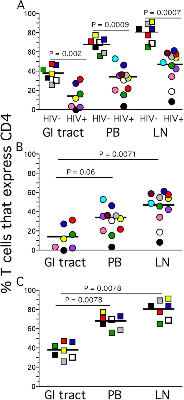
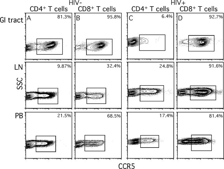
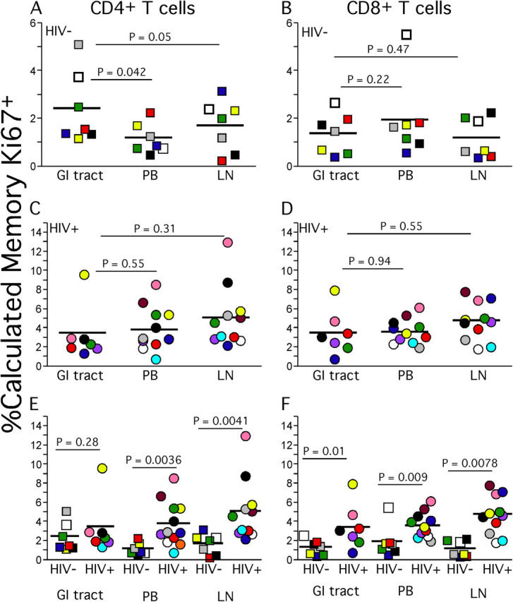
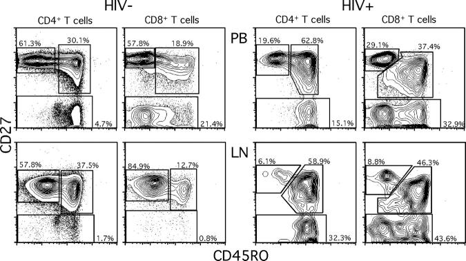
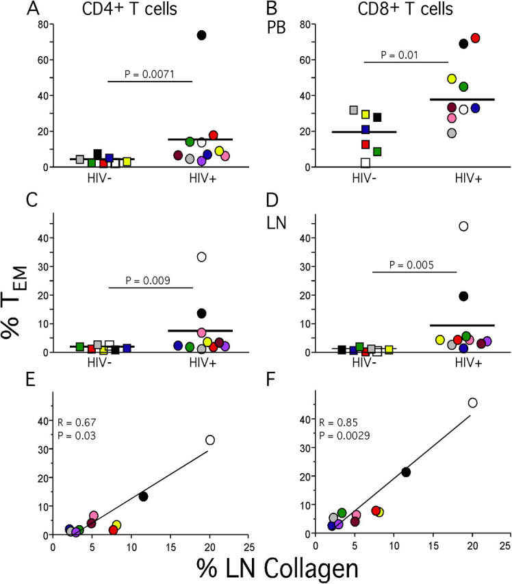
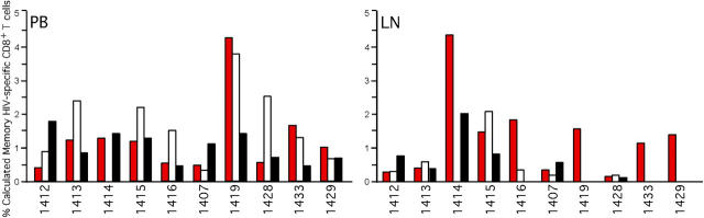
The mechanisms underlying CD4(+) T cell depletion in human immunodeficiency virus (HIV) infection are not well understood. Comparative studies of lymphoid tissues, where the vast majority of T cells reside, and peripheral blood can potentially illuminate the pathogenesis of HIV-associated disease. Here, we studied the effect of HIV infection on the activation and depletion of defined subsets of CD4(+) and CD8(+) T cells in the blood, gastrointestinal (GI) tract, and lymph node (LN). We also measured HIV-specific T cell frequencies in LNs and blood, and LN collagen deposition to define architectural changes associated with chronic inflammation. The major findings to emerge are the following: the GI tract has the most substantial CD4(+) T cell depletion at all stages of HIV disease; this depletion occurs preferentially within CCR5(+) CD4(+) T cells; HIV-associated immune activation results in abnormal accumulation of effector-type T cells within LNs; HIV-specific T cells in LNs do not account for all effector T cells; and T cell activation in LNs is associated with abnormal collagen deposition. Taken together, these findings define the nature and extent of CD4(+) T cell depletion in lymphoid tissue and point to mechanisms of profound depletion of specific T cell subsets related to elimination of CCR5(+) CD4(+) T cell targets and disruption of T cell homeostasis that accompanies chronic immune activation.


Figures

Figure 1.
Figure 1.
CD4+ T cell percentages in the GI tract, LNs, and peripheral blood. Lymphocytes from HIV+ (A and B) and HIV (A and C) individuals obtained from the blood, LNs, and GI tract were stained with anti-CD3 and anti-CD4 antibodies. The percentage of T cells that express CD4 represents lymphocyte and CD3+ gated events that stain positively with anti-CD4. p-values represent the results of Mann-Whitney statistical significance calculations between sample cohorts (HIV+ compared with HIV) or the results of Wilcoxon matched pairs for comparisons among compartments for individual cohorts.
Figure 2.
Figure 2.
Endoscopic and histological analysis of the GI tract. Endoscopic photographs from subjects 1421 (A) and 1329 (B) were obtained by passing the colonoscope past the ileo–cecal junction into the terminal ileum. Immunohistological staining for CD4 was performed by sectioning biopsies from ileal lymphoid tissue, and then hematoxylin and eosin staining followed by staining for CD4 (C and D).
Figure 3.
Figure 3.
CCR5 expression by T cells in the GI tract, LNs, and peripheral blood. GI tract, LN, and peripheral blood lymphocytes from subject 1425 (A and B, HIV) and 1428 (C and D, HIV+) were stained with anti-CD3, anti-CD4, anti-CD8, and anti-CCR5 antibodies. Plots represent lymphocyte, CD3+, and either CD4+ or CD8+ gated events. Values indicate the calculated percentage of memory CD4+ or CD8+ T cells that express CCR5 as described in Materials and Methods.
Figure 4.
Figure 4.
Ki67 expression by memory T cells in the GI tract, LNs, and peripheral blood. Lymphocytes from HIV (A, B, E, and F) and HIV+ (C–F) individuals obtained from the peripheral blood, LNs, and GI tract were stained extracellularly with anti-CD3, anti-CD4 antibodies, and anti-CD8 antibodies followed by intracellular staining with anti-Ki67. The calculated percentage of memory T cells that express Ki67 represents lymphocyte gated, CD3+, and either CD4+ or CD8+ gated events that stain positively with anti–Ki67 and were then expressed as the percentage of memory CD4+ or CD8+ T cells as described in Materials and Methods.
Figure 5.
Figure 5.
Naive, central memory, and TEM cell populations in LNs and peripheral blood. LN and peripheral blood lymphocytes from subject 1425 (HIV) and 1413 (HIV+) were stained with anti-CD4, anti-CD8, anti-CD45RO, and anti-CD27 antibodies. Plots represent lymphocyte and either CD4+ or CD8+ gated events.
Figure 6.
Figure 6.
TEM cells and collagen deposition in LNs. Percentages of LN (A and B) and peripheral blood (C and D) CD4+ and CD8+ TEM cell populations were determined based upon expression patterns of CD27 and CD45RO. Collagen deposition levels were calculated as described in Materials and Methods and were then compared with percentage of CD4+ (E) or CD8+ T cells (F) of the TEM phenotype.
Figure 7.
Figure 7.
HIV-specific CD8+ T cells in LNs and peripheral blood. Lymphocytes from HIV+ individuals obtained from peripheral blood and LNs were stimulated with overlapping HIV peptides for gag, pol, and env, and costimulatory antibodies as described in Materials and Methods. Lymphocytes were then extracellularly stained with anti-CD3, anti-CD4, and anti-CD8 antibodies followed by intracellular staining with anti–IL-2 and anti–IFN-γ antibodies. The percentage of memory CD8+ T cells that are HIV specific was determined after lymphocyte, CD3+, and CD8+ gating followed by adjustment based upon the percentage of memory CD8+ T cells for each individual as described in Materials and Methods. Bars represent the frequency of memory CD8+ T cells that respond to pol, (red bars) gag (white bars), or env (black bars).

References

    1. Dybul, M., A.S. Fauci, J.G. Bartlett, J.E. Kaplan, and A.K. Pau. 2002. Guidelines for using antiretroviral agents among HIV-infected adults and adolescents. Recommendations of the Panel on Clinical Practices for Treatment of HIV. MMWR Recomm. Rep. 51:1–55. - PubMed
    1. Mowat, A., and J. Viney. 1997. The anatomical basis of intestinal immunity. Immunol. Rev. 156:145–166. - PubMed
    1. Anton, P.A., J. Elliott, M.A. Poles, I.M. McGowan, J. Matud, L.E. Hultin, K. Grovit-Ferbas, C.R. Mackay, I.S.Y. Chen, and J.V. Giorgi. 2000. Enhanced levels of functional HIV-1 co-receptors on human mucosal T cells demonstrated using intestinal biopsy tissue. AIDS. 14:1761–1765. - PubMed
    1. Agace, W.W., A.I. Roberts, L. Wu, C. Greineder, E.C. Ebert, and C.M. Parker. 2000. Human intestinal lamina propria and intraepithelial lymphocytes express receptors specific for chemokines induced by inflammation. Eur. J. Immunol. 30:819–826. - PubMed
    1. Veazey, R.S., M. DeMaria, L.V. Chalifoux, D.E. Shvetz, D.R. Pauley, H.L. Knight, M. Rosenzweig, R.P. Johnson, R.C. Desrosiers, and A.A. Lackner. 1998. Gastrointestinal tract as a major site of CD4+ T cell depletion and viral replication in SIV infection. Science. 280:427–431. - PubMed

Publication types