. 2004 Sep 21;101(38):13762-7.
doi: 10.1073/pnas.0405883101. Epub 2004 Sep 13.

Structural basis for the interaction of Escherichia coli NusA with protein N of phage lambda

Affiliations

Structural basis for the interaction of Escherichia coli NusA with protein N of phage lambda

Irena Bonin et al. Proc Natl Acad Sci U S A. .

Abstract

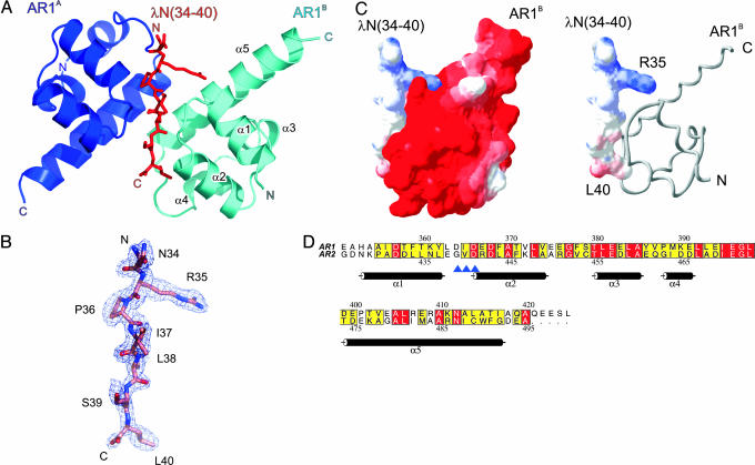
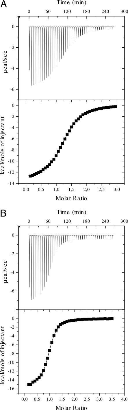
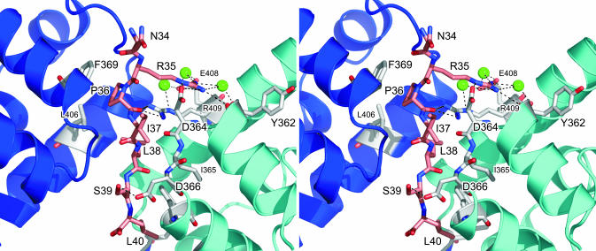
The C terminus of transcription factor NusA from Escherichia coli comprises two repeat units, which bind during antitermination to protein N from phage lambda. To delineate the structural basis of the NusA-lambdaN interaction, we attempted to crystallize the NusA C-terminal repeats in complex with a lambdaN peptide (residues 34-47). The two NusA domains became proteolytically separated during crystallization, and crystals contained two copies of the first repeat unit in contact with a single lambdaN fragment. The NusA modules employ identical regions to contact the peptide but approach the ligand from opposite sides. In contrast to the alpha-helical conformation of the lambdaN N terminus in complex with boxB RNA, residues 34-40 of lambdaN remain extended upon interaction with NusA. Mutational analyses indicated that only one of the observed NusA-lambdaN interaction modes is biologically significant, supporting an equimolar ratio of NusA and lambdaN in antitermination complexes. Solution studies indicated that additional interactions are fostered by the second NusA repeat unit, consistent with known compensatory mutations in NusA and lambdaN. Contrary to the RNA polymerase alpha subunit, lambdaN binding does not stimulate RNA interaction of NusA. The results demonstrate that lambdaN serves as a scaffold to closely oppose NusA and the mRNA in antitermination complexes.


Figures

Fig. 1.
Fig. 1.
Structure of the (NusA AR1)2–λN complex. (A) Ribbon plot of the (NusA AR1)2–λN complex (blue, AR1A; cyan, AR1B). The λN(34–40) peptide (red sticks) is sandwiched between the two AR1 α1–α2 loops. N and C termini are labeled. Structure figures were prepared with pymol (www.pymol.org). (B) Portion of the final 2FoFc electron-density map, which covers residues 34–40 of the λN peptide, contoured at the 1σ level. (C Left) Electrostatic surface potentials calculated separately for the binding partners in the physiological complex. Red, negative potential; blue, positive potential. A charge complementarity at the peptide interface can be clearly discerned. (C Right) Surface representation of the λN(34–40) peptide with NusA AR1B displayed as a gray ribbon (prepared with swisspdbviewer, http://us.expasy.org/spdbv/mainpage.html). (D) Sequence alignment of AR1 and AR2. Numbering above and below the alignment corresponds to AR1 and AR2, respectively. The background of identical residues is red, and the background of conserved amino acids is yellow. Secondary structure elements as found for AR1 are indicated. Blue arrows indicate residues of AR1 (D364–D366), which align in a β-like arrangement with I37–S39 of the peptide. Prepared with alscript (36).
Fig. 2.
Fig. 2.
Probing of the NusA–λN interactions by ITC. (A) Reaction of NusA AR1–AR2 with λN(31–43). (B) Reaction of NusA AR1–AR2 with λN(34–47). The negative peaks indicate an exothermic reaction. The area under each peak represents the heat released after an injection of the peptide into the solution of NusA. (Lower) Binding isotherms obtained by plotting peak areas against the molar ratio of λN peptide to NusA. The lines represent the best-fit curves obtained from least-squares regression analyses assuming a one-site binding model.
Fig. 3.
Fig. 3.
Stereoview detailing the AR1–λN(34–40) interaction. The backbone ribbons of the AR1A and AR1B molecules are in blue and cyan, respectively. Residues interacting specifically with the peptide are shown as sticks and are color-coded by atom type (gray, carbon; red, oxygen; blue, nitrogen). The λN peptide is drawn as a stick figure (pink, carbon). Three water molecules, which are mediating interactions, are shown as green spheres. Hydrogen bonds or salt bridges discussed in the text are indicated as dashed lines.

References

    1. Greenblatt, J., Mah, T. F., Legault, P., Mogridge, J., Li, J. & Kay, L. E. (1998) Cold Spring Harbor Symp. Quant. Biol. 63, 327–336. - PubMed
    1. Nudler, E. & Gottesman, M. E. (2002) Genes Cells 7, 755–768. - PubMed
    1. Mogridge, J., Mah, T. F. & Greenblatt, J. (1995) Genes Dev. 9, 2831–2845. - PubMed
    1. Mogridge, J., Mah, T. F. & Greenblatt, J. (1998) J. Biol. Chem. 273, 4143–4148. - PubMed
    1. Van Gilst, M. R. & von Hippel, P. H. (1997) J. Mol. Biol. 274, 160–173. - PubMed

Publication types

MeSH terms

Associated data