

Cloning and characterization of murine 1-acyl-sn-glycerol 3-phosphate acyltransferases and their regulation by PPARalpha in murine heart
- PMID: 15367102
- PMCID: PMC1134718
- DOI: 10.1042/BJ20041348
Cloning and characterization of murine 1-acyl-sn-glycerol 3-phosphate acyltransferases and their regulation by PPARalpha in murine heart
Abstract
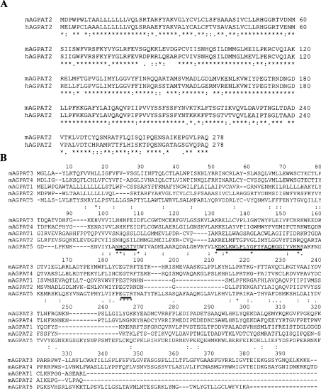
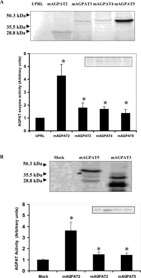
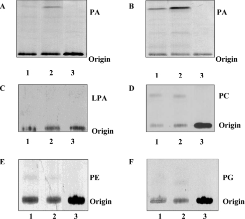
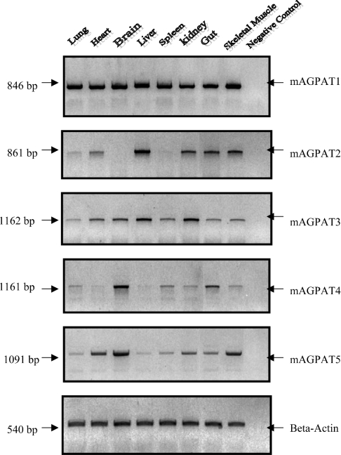
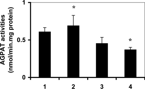
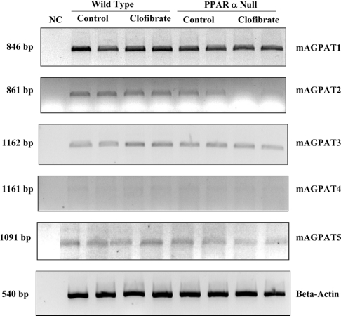
AGPAT (1-acyl-sn-glycerol 3-phosphate acyltransferase) exists in at least five isoforms in humans, termed as AGPAT1, AGPAT2, AGPAT3, AGPAT4 and AGPAT5. Although they catalyse the same biochemical reaction, their relative function, tissue expression and regulation are poorly understood. Linkage studies in humans have revealed that AGPAT2 contributes to glycerolipid synthesis and plays an important role in regulating lipid metabolism. We report the molecular cloning, tissue distribution, and enzyme characterization of mAGPATs (murine AGPATs) and regulation of cardiac mAGPATs by PPARalpha (peroxisome-proliferator-activated receptor alpha). mAGPATs demonstrated differential tissue expression profiles: mAGPAT1 and mAGPAT3 were ubiquitously expressed in most tissues, whereas mAGPAT2, mAGPAT4 and mAGPAT5 were expressed in a tissue-specific manner. mAGPAT2 expressed in in vitro transcription and translation reactions and in transfected COS-1 cells exhibited specificity for 1-acyl-sn-glycerol 3-phosphate. When amino acid sequences of five mAGPATs were compared, three highly conserved motifs were identified, including one novel motif/pattern KX2LX6GX12R. Cardiac mAGPAT activities were 25% lower (P<0.05) in PPARalpha null mice compared with wild-type. In addition, cardiac mAGPAT activities were 50% lower (P<0.05) in PPARalpha null mice fed clofibrate compared with clofibrate fed wild-type animals. This modulation of AGPAT activity was accompanied by significant enhancement/reduction of the mRNA levels of mAGPAT3/mAGPAT2 respectively. Finally, mRNA expression of cardiac mAGPAT3 appeared to be regulated by PPARalpha activation. We conclude that cardiac mAGPAT activity may be regulated by both the composition of mAGPAT isoforms and the levels of each isoform.
Figures
Similar articles
-
cDNA cloning and expression of murine 1-acyl-sn-glycerol-3-phosphate acyltransferase.Biochem Biophys Res Commun. 1997 Aug 28;237(3):663-6. doi: 10.1006/bbrc.1997.7214. Biochem Biophys Res Commun. 1997. PMID: 9299423
-
Expression and regulation of 1-acyl-sn-glycerol- 3-phosphate acyltransferases in the epidermis.J Lipid Res. 2005 Nov;46(11):2448-57. doi: 10.1194/jlr.M500258-JLR200. Epub 2005 Sep 8. J Lipid Res. 2005. PMID: 16150824
-
Functional characterization of human 1-acylglycerol-3-phosphate acyltransferase isoform 8: cloning, tissue distribution, gene structure, and enzymatic activity.Arch Biochem Biophys. 2006 May 15;449(1-2):64-76. doi: 10.1016/j.abb.2006.03.014. Epub 2006 Mar 29. Arch Biochem Biophys. 2006. PMID: 16620771
-
Lysophospholipid acyltransferases: 1-acylglycerol-3-phosphate O-acyltransferases. From discovery to disease.Curr Opin Lipidol. 2012 Aug;23(4):290-302. doi: 10.1097/MOL.0b013e328354fcf4. Curr Opin Lipidol. 2012. PMID: 22777291 Review.
-
The structure and functions of human lysophosphatidic acid acyltransferases.Front Biosci. 2001 Aug 1;6:D944-53. doi: 10.2741/leung. Front Biosci. 2001. PMID: 11487472 Review.
Cited by
-
Transcriptional programming of lipid and amino acid metabolism by the skeletal muscle circadian clock.PLoS Biol. 2018 Aug 10;16(8):e2005886. doi: 10.1371/journal.pbio.2005886. eCollection 2018 Aug. PLoS Biol. 2018. PMID: 30096135 Free PMC article.
-
Dengue virus reduces AGPAT1 expression to alter phospholipids and enhance infection in Aedes aegypti.PLoS Pathog. 2019 Dec 9;15(12):e1008199. doi: 10.1371/journal.ppat.1008199. eCollection 2019 Dec. PLoS Pathog. 2019. PMID: 31815960 Free PMC article.
-
Recent progress on acyl CoA: lysophospholipid acyltransferase research.J Lipid Res. 2009 Apr;50 Suppl(Suppl):S46-51. doi: 10.1194/jlr.R800035-JLR200. Epub 2008 Oct 17. J Lipid Res. 2009. PMID: 18931347 Free PMC article. Review.
-
Mechanotransduction and the regulation of mTORC1 signaling in skeletal muscle.Int J Biochem Cell Biol. 2011 Sep;43(9):1267-76. doi: 10.1016/j.biocel.2011.05.007. Epub 2011 May 19. Int J Biochem Cell Biol. 2011. PMID: 21621634 Free PMC article. Review.
-
LPS and proinflammatory cytokines decrease lipin-1 in mouse adipose tissue and 3T3-L1 adipocytes.Am J Physiol Endocrinol Metab. 2008 Dec;295(6):E1502-9. doi: 10.1152/ajpendo.90323.2008. Epub 2008 Oct 21. Am J Physiol Endocrinol Metab. 2008. PMID: 18940942 Free PMC article.
References
-
- Vance D. E. Phospholipid biosynthesis in eukaryotes. In: Vance D. E., Vance J., editors. Biochemistry of Lipids, Lipoproteins and Membranes. 4th edn. Amsterdam: Elsevier Science; 2002. pp. 205–231.
-
- Bursten S. L., Harris W. E., Bomsztyk K., Lovett D. Interleukin-1 rapidly stimulates lysophosphatidate acyltransferase and phosphatidate phosphohydrolase activities in human mesangial cells. J. Biol. Chem. 1991;266:20732–20743. - PubMed
-
- English D., Garcia J. G., Brindley D. N. Platelet-released phospholipids link haemostasis and angiogenesis. Cardiovasc. Res. 2001;49:588–599. - PubMed
-
- Hla T., Lee M. J., Ancellin N., Paik J. H., Kluk M. J. Lysophospholipids–receptor revelations. Science. 2001;294:1875–1878. - PubMed
-
- McIntyre T. M., Pontsler A. V., Silva A. R., St Hilire A., Su Y., Hinshow J. C., Zimmerman G. A., Hama K., Aoki J., Arai H., et al. Identification of an intracellular receptor for lysophosphatidic acid (LPA): LPA is a transcellular PPARgamma agonist. Proc. Natl. Acad. Sci. U.S.A. 2003;100:131–136. - PMC - PubMed
Publication types
MeSH terms
Substances
LinkOut - more resources
Full Text Sources
Other Literature Sources
Molecular Biology Databases