. 2005 Jan 15;385(Pt 2):485-91.
doi: 10.1042/BJ20040694.

5-aminoimidazole-4-carboxamide riboside (AICAR) enhances GLUT2-dependent jejunal glucose transport: a possible role for AMPK

Affiliations

5-aminoimidazole-4-carboxamide riboside (AICAR) enhances GLUT2-dependent jejunal glucose transport: a possible role for AMPK

John Walker et al. Biochem J. .

Abstract

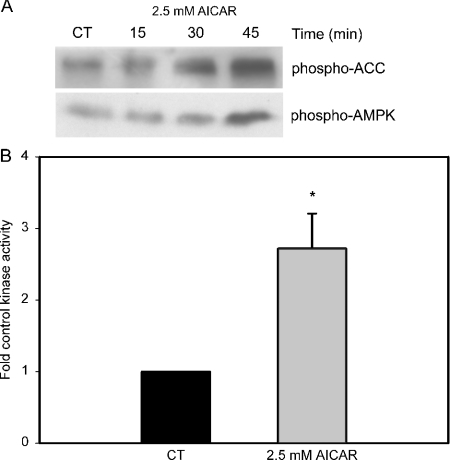
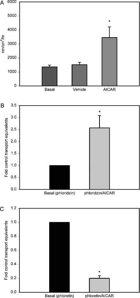
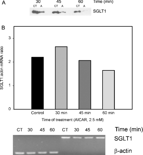
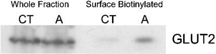
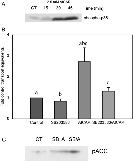
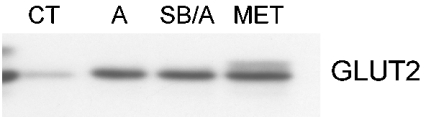
AMPK (AMP-activated protein kinase) is a key sensor of energy status within the cell. Activated by an increase in the AMP/ATP ratio, AMPK acts to limit cellular energy depletion by down-regulating selective ATP-dependent processes. The purpose of the present study was to determine the role of AMPK in regulating intestinal glucose transport. [3H]3-O-methyl glucose fluxes were measured in murine jejunum in the presence and absence of the AMPK activators AICAR (5-aminoimidazole-4-carboxamide riboside) and metformin and the p38 inhibitor, SB203580. To differentiate between a sodium-coupled (SGLT1) and diffusive (GLUT2) route of entry, fluxes were measured in the presence of the SGLT1 and GLUT2 inhibitors phloridzin and phloretin. Glucose transporter mRNA levels were measured by reverse transcriptase-PCR, and localization by Western blotting. Surface-expressed GLUT2 was assessed by luminal biotinylation. Activation of p38 mitogen-activated protein kinase was analysed by Western blotting. We found that treatment of jejunal tissue with AICAR resulted in enhanced net glucose uptake and was associated with phosphorylation of p38 mitogen-activated protein kinase. Inhibition of p38 abrogated the stimulation of AICAR-stimulated glucose uptake. Phloretin abolished the AICAR-mediated increase in glucose flux, whereas phloridzin had no effect, suggesting the involvement of GLUT2. In addition, AICAR decreased total protein levels of SGLT1, concurrently increasing levels of GLUT2 in the brush-border membrane. The anti-diabetic drug metformin, a known activator of AMPK, also induced the localization of GLUT2 to the luminal surface. We conclude that the activation of AMPK results in an up-regulation of non-energy requiring glucose uptake by GLUT2 and a concurrent down-regulation of sodium-dependent glucose transport.


Figures

Figure 1
Figure 1. Time-dependent phosphorylation and activation of AMPK and phosphorylation of ACC with treatment by AICAR
(A) Western blot of isolated jejunum incubated with 2.5 mM AICAR at 37 °C in oxygenated normal Ringer's solution for the time period indicated. In both experiments, the control group was jejunal tissue treated with vehicle (0.25% DMSO) for the maximum incubation period represented in the AICAR group. After treatment, mucosal scrapings were analysed by Western blot for the phosphorylation state of the indicated targets. (B) AMPK activity assay of isolated jejunum incubated with 2.5 mM AICAR (30 min treatment). The control group was jejunal tissue treated with vehicle (0.25% DMSO). Error bars represent S.E.M. Significance was determined by unpaired Student's t test (*P<0.05, n=5). CT, control.
Figure 2
Figure 2. Uptake of 3-O-methyl glucose from isolated jejunal tissue
(A) Jejunal tissue was mounted in Lucite chambers exposing mucosal and serosal surfaces to 10 ml of oxygenated Krebs buffer. Net directional 3-O-methyl glucose flux from mucosal-to-serosal surface was determined by measuring four consecutive 5 min fluxes before the addition of AICAR (2.5 mM) and four 5 min fluxes after the addition of the AMPK activator. (B) Values represent net directional flux as measured in (A), except in the presence of the SGLT1 inhibitor phloridzin (1 mM). (C) Values represent net directional flux as measured in (A), except in the presence of the GLUT2 inhibitor phloretin (1 mM). Error bars represent S.E.M. Significance was determined by ANOVA (A) or unpaired Student's t test (B, C), and groups found to be statistically distinct from control are denoted by asterisk (*P<0.05, n≥5 animals per group).
Figure 3
Figure 3. Alterations in SGLT1 transporter and mRNA levels in response to AICAR incubation
(A) Western blot of isolated jejunum incubated with 2.5 mM AICAR at 37 °C in oxygenated normal Ringer's solution for the time period indicated. The control group was jejunal tissue treated with vehicle (0.25% DMSO) for the respective incubation period represented in the AICAR group. After treatment, mucosal scrapings were analysed by Western blot for total cellular levels of SGLT1. (B) RT–PCR experiment examining the level of SGLT1 mRNA following incubation of isolated jejunal tissue with 2.5 mM AICAR at 37 °C in oxygenated normal Ringer's solution for the time periods indicated. Values are expressed as the ratio of amplified SGLT1/β-actin cDNA. Differences indicated between treatment groups were not found to be statistically different. CT, control; A, AICAR.
Figure 4
Figure 4. Alterations in total cellular and brush-border-localized GLUT2 in response to AICAR incubation
Western blot of isolated jejunum incubated with 2.5 mM AICAR at 37 °C in oxygenated normal Ringer's solution for 45 min. Luminal proteins were labelled with biotin and examined by Western blotting for alterations in GLUT2 protein. Those lanes labelled ‘Whole Fraction’ are representative of the tissue preparation before the extraction of the biotinylated proteins, and are taken as indicative of total cellular levels of GLUT2. Lanes labelled ‘Surface Biotinylated’ are representative of isolated biotinylated proteins, and are representative of surface-expressed GLUT2. The control group was jejunal tissue treated with vehicle (0.25% DMSO) for an incubation period equivalent to the treatment groups. CT, control; A, AICAR.
Figure 5
Figure 5. p38 MAPK phosphorylation is required for AICAR-stimulated glucose transport
(A) Western blot of isolated jejunum incubated with 2.5 mM AICAR at 37 °C in oxygenated normal Ringer's solution for the time periods indicated. The control group was jejunal tissue treated with vehicle (0.25% DMSO) for the maximum incubation period represented in the AICAR group. Mucosal scrapings were analysed for phospho-p38 MAPK. (B) Net directional 3-O-methyl glucose flux from mucosal-to-serosal before, and after the addition of AICAR (2.5 mM). Where treatment involved the p38 inhibitor SB203580 (20 μM) the jejunum was perfused in vivo for a period of 30 min before the analysis of in vitro glucose transport as described in all other transport experiments. Error bars represent S.E.M. Significance was determined by ANOVA; groups found to be statistically distinct from one another share the same letter (a and b, P<0.01; c, P<0.05, n≥5 animals per group). (C) Western blot of isolated jejunum incubated with 2.5 μM AICAR and/or SB203580 (20 μM) showing phosphorylation of the AMPK substrate ACC (phospho-ACC). The control group was jejunal tissue treated with vehicle (0.25% DMSO) for a period equivalent to the treatment groups. CT, control; A, AICAR; SB, SB203580.
Figure 6
Figure 6. AICAR-dependent GLUT2 translocation to the apical surface is not dependent on p38 MAPK
Western blot of isolated jejunal tissue incubated with 2.5 mM AICAR (30 min) at 37 °C in oxygenated normal Ringer's solution with and without 30 min pretreatment with the p38 inhibitor SB203580 (20 μM). Treatment by the AMPK activator metformin (5 mM) was performed as with AICAR. The control group was jejunal tissue treated with vehicle (0.25% DMSO) for an incubation period equivalent to the treatment groups. Lanes are representative of isolated biotinylated proteins and are representative of surface-expressed GLUT2. CT, control; A, AICAR; SB, SB203580; MET, metformin.

Similar articles

Cited by

References

    1. Hardie D. G., Scott J. W., Pan D. A., Hudson E. R. Management of cellular energy by the AMP-activated protein kinase system. FEBS Lett. 2003;546:113–120. - PubMed
    1. Carling D., Clarke P. R., Zammit V. A., Hardie D. G. Purification and characterization of the AMP-activated protein kinase. Copurification of acetyl-CoA carboxylase kinase and 3-hydroxy-3-methylglutaryl-CoA reductase kinase activities. Eur. J. Biochem. 1989;186:129–136. - PubMed
    1. Stein S. C., Woods A., Jones N. A., Davison M. D., Carling D. The regulation of AMP-activated protein kinase by phosphorylation. Biochem. J. 2000;345:437–443. - PMC - PubMed
    1. Kemp B. E., Stapleton D., Campbell D. J., Chen Z. P., Murthy S., Walter M., Gupta A., Adams J. J., Katsis F., van Denderen B., et al. AMP-activated protein kinase, super metabolic regulator. Biochem. Soc. Trans. 2003;31:162–168. - PubMed
    1. Carling D. AMPK. Curr. Biol. 2004;14:R220. - PubMed

Publication types

MeSH terms