. 2004 Oct;78(19):10221-9.
doi: 10.1128/JVI.78.19.10221-10229.2004.

Human cytomegalovirus TRS1 protein is required for efficient assembly of DNA-containing capsids

Affiliations

Human cytomegalovirus TRS1 protein is required for efficient assembly of DNA-containing capsids

Joan E Adamo et al. J Virol. 2004 Oct.

Abstract

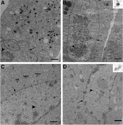
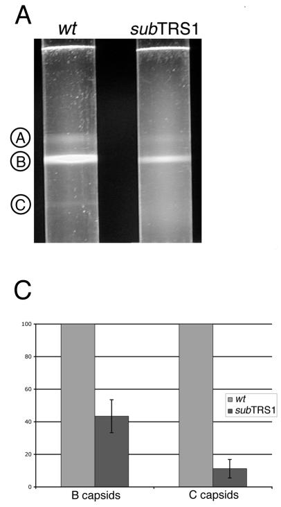
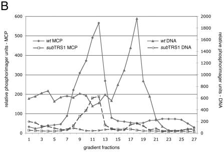
The human cytomegalovirus tegument protein, pTRS1, appears to function at several discrete stages of the virus replication cycle. We previously demonstrated that pTRS1 acts during the late phase of infection to facilitate the production of infectious virions. We now have more precisely identified the late pTRS1 function by further study of a mutant virus lacking the TRS1 region, ADsubTRS1. We observed a significant reduction in the production of capsids, especially DNA-containing C-capsids, in mutant virus-infected cells. ADsubTRS1 exhibited normal cleavage of DNA concatemers, so the defect in C-capsid production must occur after DNA cleavage and before DNA is stably inserted into a capsid. Further, the normal virus-induced morphological reorganization of the nucleus did not occur after infection with the pTRS1-deficient mutant.


Figures

FIG. 1.
FIG. 1.
Microarray analysis of the ADwt (A) and ADsubTRS1 (B) mutant reveals similar patterns of HCMV gene expression in infected fibroblasts. DNA arrays were probed with cDNA prepared from fibroblasts infected at an MOI of 0.5 PFU/cell and harvested at 72 h postinfection. The top three panels in each section have cDNAs that span the entire HCMV genome along with several controls. The bottom panel in each section contains a set of control cellular genes.
FIG.2.
FIG.2.
Immunofluorescence analysis of the intracellular localization of HCMV proteins involved in capsid formation and DNA replication after infection with ADwt (A, C, E, G, I, and K) or ADsubTRS1 (B, D, F, H, J, and L). Fibroblasts were fixed at 72 h postinfection and probed with MAbs specific to the pUL86 major capsid protein (A and B), pUL85 minor capsid protein (C and D), pUL48.5 smallest capsid protein (E and F), pUL112/113 replication compartment protein (G and H), pUL44 polymerase accessory proteins (I and J), and pUL98 virus-coded DNase (K and L). The left panel in each pair shows the antibody-specific signal alone (red), and in the right panel of each pair the DNA is counterstained with DAPI (blue). Bars, 10 μm.
FIG. 3.
FIG. 3.
The ADsubTRS1 mutant causes a decrease in capsid production and a disruption of viral replication centers. Thin-section transmission electron microscopy of fibroblasts infected at an MOI of 0.5 PFU/cell and fixed at 72 h postinfection. Fibroblasts were infected with ADwt (A and B) or ADsubTRS1 (C and D). The nuclear envelope is indicated by arrows, and the arrowheads point to capsids. NIEPs, virions, and DBs are labeled in the cytoplasm (A and C). All images (except for higher magnification insets) are taken at the same magnification. Bars, 1 μm.
FIG. 4.
FIG. 4.
Rate velocity gradient separation of HCMV nucleocapsids. Lysates from ADwt- or ADsubTRS1-infected fibroblasts were layered onto 20 to 65% sucrose gradients. (A) Illumination from the top revealed three light-scattering bands in ADwt, indicated by circled letters. A-capsids are empty; B-capsids are precursors that contain the scaffolding protein, which is then released from the capsid to allow DNA insertion and the maturation to the C-capsid form. (B) Profile of gradient fractions probed for the presence of capsid proteins or HCMV DNA. This representative sample shows the distribution of both the major capsid protein (on the left axis) and viral DNA (on the right axis) along the fractions of the rate velocity gradient. (C) When the ADsubTRS1 mutant is compared to ADwt, the reduction in capsid numbers is different for the two different capsid types analyzed. Quantitation for both the B and C peaks shows the fold decrease for ADsubTRS1 compared to the ADwt strain.
FIG. 4.
FIG. 4.
Rate velocity gradient separation of HCMV nucleocapsids. Lysates from ADwt- or ADsubTRS1-infected fibroblasts were layered onto 20 to 65% sucrose gradients. (A) Illumination from the top revealed three light-scattering bands in ADwt, indicated by circled letters. A-capsids are empty; B-capsids are precursors that contain the scaffolding protein, which is then released from the capsid to allow DNA insertion and the maturation to the C-capsid form. (B) Profile of gradient fractions probed for the presence of capsid proteins or HCMV DNA. This representative sample shows the distribution of both the major capsid protein (on the left axis) and viral DNA (on the right axis) along the fractions of the rate velocity gradient. (C) When the ADsubTRS1 mutant is compared to ADwt, the reduction in capsid numbers is different for the two different capsid types analyzed. Quantitation for both the B and C peaks shows the fold decrease for ADsubTRS1 compared to the ADwt strain.
FIG. 5.
FIG. 5.
Processing of newly replicated viral DNA. Fibroblasts were infected with either ADwt or ADsubTRS1 virus at an MOI of 0.5 PFU/cell, and the total DNA was prepared at the indicated times. (A) Shows the relevant fragments of an HCMV genome digested with HpaI. (B) A representative Southern blot with a 32P-labeled probe specific for RL3. Purified cell-free viral DNA was used as a control for size. The indicated fused and free fragments were quantified by using a phosphorimager.
FIG. 6.
FIG. 6.
FISH of fibroblasts fixed infected with ADwt (A and B) or ADsubTRS1 (C and D) and then fixed at 24 h (A and C) or 96 h postinfection (B and D). HCMV DNA was visualized by FISH analysis and appears in green. Cellular DNA is counterstained with propidium iodide and appears in red.

References

    1. AbuBakar, S., W. W. Au, M. S. Legator, and T. Albrecht. 1988. Induction of chromosome aberrations and mitotic arrest by cytomegalovirus in human cells. Environ. Mol. Mutagen. 12:409-420. - PubMed
    1. Ali, M. A., B. Forghani, and E. M. Cantin. 1996. Characterization of an essential HSV-1 protein encoded by the UL25 gene reported to be involved in virus penetration and capsid assembly. Virology 216:278-283. - PubMed
    1. Blankenship, C. A., and T. Shenk. 2002. Mutant human cytomegalovirus lacking the immediate-early TRS1 coding region exhibits a late defect. J. Virol. 76:12290-12299. - PMC - PubMed
    1. Bogner, E., K. Radsak, and M. F. Stinski. 1998. The gene product of human cytomegalovirus open reading frame UL56 binds the pac motif and has specific nuclease activity. J. Virol. 72:2259-2264. - PMC - PubMed
    1. Child, S. J., M. Hakki, K. L. De Niro, and A. P. Geballe. 2004. Evasion of cellular antiviral responses by human cytomegalovirus TRS1 and IRS1. J. Virol. 78:197-205. - PMC - PubMed

Publication types

MeSH terms

LinkOut - more resources